| کد مقاله | کد نشریه | سال انتشار | مقاله انگلیسی | نسخه تمام متن |
|---|---|---|---|---|
| 1761191 | 1019637 | 2018 | 9 صفحه PDF | دانلود رایگان |
عنوان انگلیسی مقاله ISI
Classification of the Thyroid Nodules Based on Characteristic Sonographic Textural Feature and Correlated Histopathology Using Hierarchical Support Vector Machines
دانلود مقاله + سفارش ترجمه
دانلود مقاله ISI انگلیسی
رایگان برای ایرانیان
کلمات کلیدی
موضوعات مرتبط
مهندسی و علوم پایه
فیزیک و نجوم
آکوستیک و فرا صوت
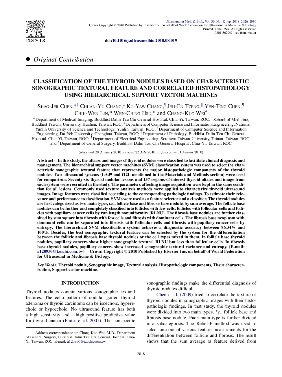
پیش نمایش صفحه اول مقاله

چکیده انگلیسی
In this study, the ultrasound images of thyroid nodules were classified to facilitate clinical diagnosis and management. The hierarchical support vector machines (SVM) classification system was used to select the characteristic sonographic textural feature that represents the major histopathologic components of the thyroid nodules. Two ultrasound systems (LA39 and i12L mentioned in the Materials and Methods section) were used for comparison. Seventy-six thyroid nodular lesions and 157 regions-of-interest thyroid ultrasound image from each system were recruited in the study. The parameters affecting image acquisition were kept in the same condition for all lesions. Commonly used texture analysis methods were applied to characterize thyroid ultrasound images. Image features were classified according to the corresponding pathologic findings. To estimate their relevance and performance to classification, SVMs were used as a feature selector and a classifier. The thyroid nodules are first categorized as two main types, i.e., follicle base and fibrosis base nodule, by sum average. The follicle base nodules can be further and completely classified into follicles with few cells, follicles with follicular cells and follicles with papillary cancer cells by run length nonuniformity (RLNU). The fibrosis base nodules are further classified by sum square into fibrosis with few cells and fibrosis with dominant cells. The fibrosis base neoplasm with dominant cells can be separated into fibrosis with follicular cells and fibrosis with papillary cancer cells by entropy. The hierarchical SVM classification system achieves a diagnostic accuracy between 96.34% and 100%. Besides, the best sonographic textural feature can be selected by the system for the differentiation between the follicle and fibrosis base thyroid nodules or the cell types mixed in them. In follicle base thyroid nodules, papillary cancers show higher sonographic textural RLNU but less than follicular cells. In fibrosis base thyroid nodules, papillary cancers show increased sonographic textural variance and entropy. (E-mail: a120930@tzuchi.com.tw)
ناشر
Database: Elsevier - ScienceDirect (ساینس دایرکت)
Journal: Ultrasound in Medicine & Biology - Volume 36, Issue 12, December 2010, Pages 2018-2026
Journal: Ultrasound in Medicine & Biology - Volume 36, Issue 12, December 2010, Pages 2018-2026
نویسندگان
Shao-Jer Chen, Chuan-Yu Chang, Ku-Yaw Chang, Jeh-En Tzeng, Yen-Ting Chen, Chih-Wen Lin, Wen-Ching Hsu, Chang-Kuo Wei,