| کد مقاله | کد نشریه | سال انتشار | مقاله انگلیسی | نسخه تمام متن |
|---|---|---|---|---|
| 5023300 | 1470248 | 2017 | 18 صفحه PDF | دانلود رایگان |
- Volumetric compressive characteristics of a gellan-gum bioactive-glass scaffold for bone tissue engineering are assessed.
- Deforming microstructure of the scaffold is observed during on-the-fly X-ray micro-tomography under loading.
- In-house micro-tomographical setup equipped with loading device is used to develop voxel model of deforming microstructure.
- High-resolution displacement and strain fields are evaluated using custom digital volume correlation method.
- Reliability and ability of proposed method to capture the deformation of microstructure with high confidence are discussed.
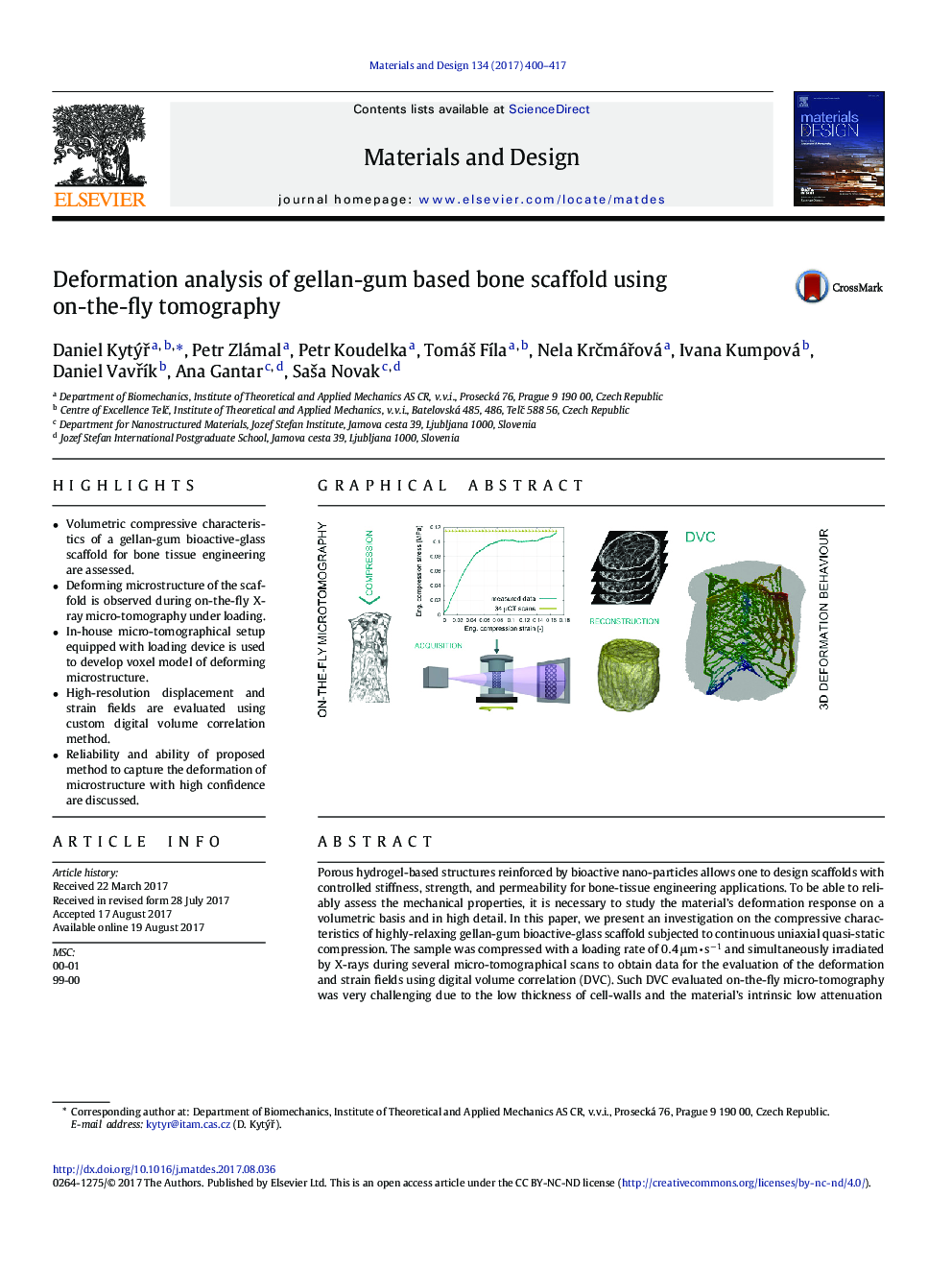
Porous hydrogel-based structures reinforced by bioactive nano-particles allows one to design scaffolds with controlled stiffness, strength, and permeability for bone-tissue engineering applications. To be able to reliably assess the mechanical properties, it is necessary to study the material's deformation response on a volumetric basis and in high detail. In this paper, we present an investigation on the compressive characteristics of highly-relaxing gellan-gum bioactive-glass scaffold subjected to continuous uniaxial quasi-static compression. The sample was compressed with a loading rate of 0.4 μmâ s â1 and simultaneously irradiated by X-rays during several micro-tomographical scans to obtain data for the evaluation of the deformation and strain fields using digital volume correlation (DVC). Such DVC evaluated on-the-fly micro-tomography was very challenging due to the low thickness of cell-walls and the material's intrinsic low attenuation of X-rays. Thus, we employed loading and tomographical devices equipped with a single-photon counting detector coupled with a DVC procedure, all developed in-house. From the acquired 34 tomographical scans, high-resolution voxel models with a resolution of 29.77 μm were developed and subjected to DVC to obtain detailed deformation and strain fields of the material. It is shown that the presented method is suitable for the precise determination of the deformation response of the predominantly organic material developed as a biocompatible, bioresorbable bone scaffold.
Graphical Abstract379
Journal: Materials & Design - Volume 134, 15 November 2017, Pages 400-417
