| کد مقاله | کد نشریه | سال انتشار | مقاله انگلیسی | نسخه تمام متن |
|---|---|---|---|---|
| 5718560 | 1607135 | 2017 | 6 صفحه PDF | دانلود رایگان |

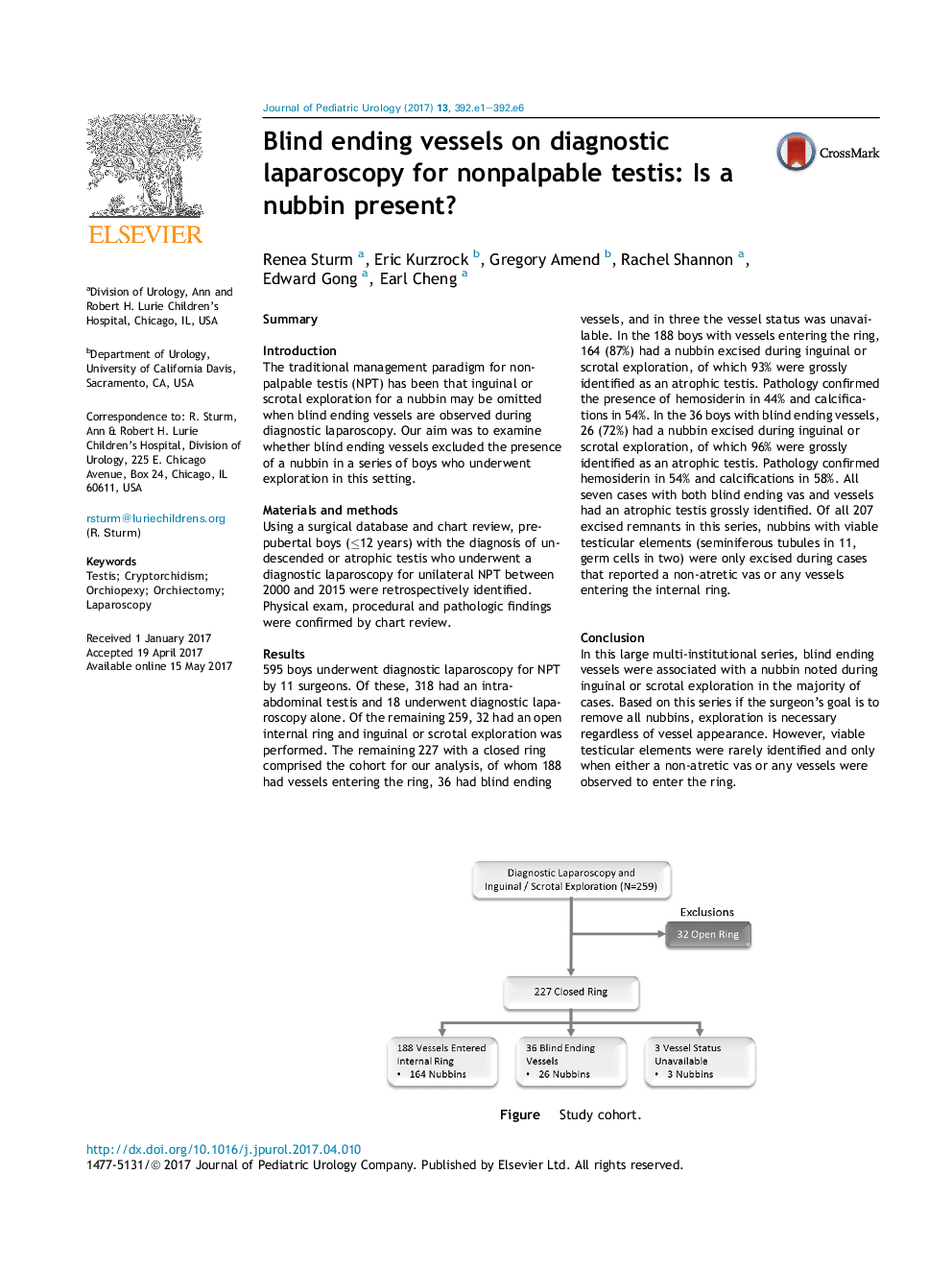
SummaryIntroductionThe traditional management paradigm for nonpalpable testis (NPT) has been that inguinal or scrotal exploration for a nubbin may be omitted when blind ending vessels are observed during diagnostic laparoscopy. Our aim was to examine whether blind ending vessels excluded the presence of a nubbin in a series of boys who underwent exploration in this setting.Materials and methodsUsing a surgical database and chart review, pre-pubertal boys (â¤12 years) with the diagnosis of undescended or atrophic testis who underwent a diagnostic laparoscopy for unilateral NPT between 2000 and 2015 were retrospectively identified. Physical exam, procedural and pathologic findings were confirmed by chart review.Results595 boys underwent diagnostic laparoscopy for NPT by 11 surgeons. Of these, 318 had an intra-abdominal testis and 18 underwent diagnostic laparoscopy alone. Of the remaining 259, 32 had an open internal ring and inguinal or scrotal exploration was performed. The remaining 227 with a closed ring comprised the cohort for our analysis, of whom 188 had vessels entering the ring, 36 had blind ending vessels, and in three the vessel status was unavailable. In the 188 boys with vessels entering the ring, 164 (87%) had a nubbin excised during inguinal or scrotal exploration, of which 93% were grossly identified as an atrophic testis. Pathology confirmed the presence of hemosiderin in 44% and calcifications in 54%. In the 36 boys with blind ending vessels, 26 (72%) had a nubbin excised during inguinal or scrotal exploration, of which 96% were grossly identified as an atrophic testis. Pathology confirmed hemosiderin in 54% and calcifications in 58%. All seven cases with both blind ending vas and vessels had an atrophic testis grossly identified. Of all 207 excised remnants in this series, nubbins with viable testicular elements (seminiferous tubules in 11, germ cells in two) were only excised during cases that reported a non-atretic vas or any vessels entering the internal ring.ConclusionIn this large multi-institutional series, blind ending vessels were associated with a nubbin noted during inguinal or scrotal exploration in the majority of cases. Based on this series if the surgeon's goal is to remove all nubbins, exploration is necessary regardless of vessel appearance. However, viable testicular elements were rarely identified and only when either a non-atretic vas or any vessels were observed to enter the ring.150Figure. Study cohort.
Journal: Journal of Pediatric Urology - Volume 13, Issue 4, August 2017, Pages 392.e1-392.e6