| کد مقاله | کد نشریه | سال انتشار | مقاله انگلیسی | نسخه تمام متن |
|---|---|---|---|---|
| 8504224 | 1554330 | 2018 | 5 صفحه PDF | دانلود رایگان |
عنوان انگلیسی مقاله ISI
Application of colour Doppler and three-dimensional (3D) ultrasonography for visualization of mammary gland structures in goats
دانلود مقاله + سفارش ترجمه
دانلود مقاله ISI انگلیسی
رایگان برای ایرانیان
موضوعات مرتبط
علوم زیستی و بیوفناوری
علوم کشاورزی و بیولوژیک
علوم دامی و جانورشناسی
پیش نمایش صفحه اول مقاله

چکیده انگلیسی
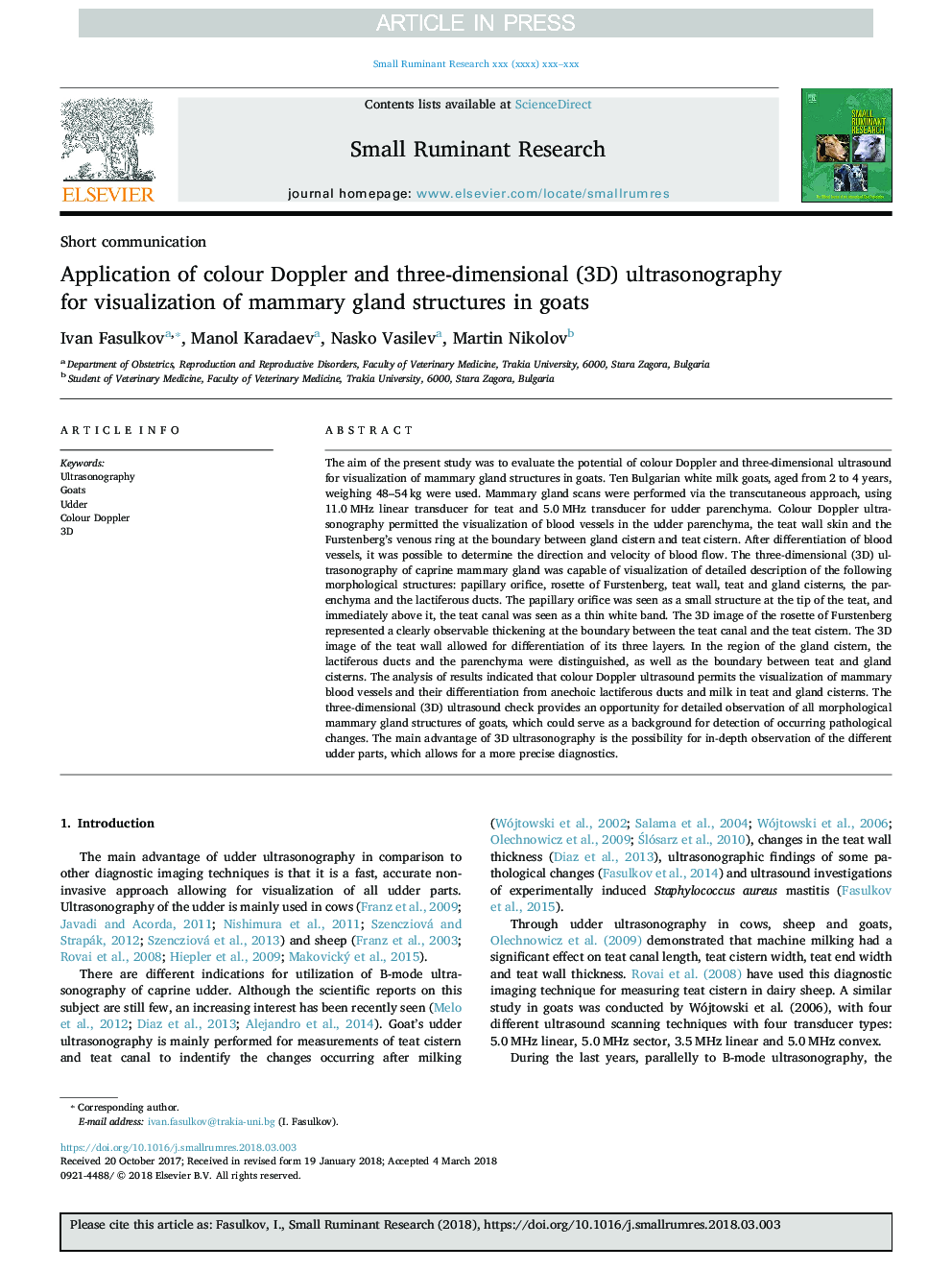
The aim of the present study was to evaluate the potential of colour Doppler and three-dimensional ultrasound for visualization of mammary gland structures in goats. Ten Bulgarian white milk goats, aged from 2 to 4 years, weighing 48-54â¯kg were used. Mammary gland scans were performed via the transcutaneous approach, using 11.0â¯MHz linear transducer for teat and 5.0â¯MHz transducer for udder parenchyma. Colour Doppler ultrasonography permitted the visualization of blood vessels in the udder parenchyma, the teat wall skin and the Furstenberg's venous ring at the boundary between gland cistern and teat cistern. After differentiation of blood vessels, it was possible to determine the direction and velocity of blood flow. The three-dimensional (3D) ultrasonography of caprine mammary gland was capable of visualization of detailed description of the following morphological structures: papillary orifice, rosette of Furstenberg, teat wall, teat and gland cisterns, the parenchyma and the lactiferous ducts. The papillary orifice was seen as a small structure at the tip of the teat, and immediately above it, the teat canal was seen as a thin white band. The 3D image of the rosette of Furstenberg represented a clearly observable thickening at the boundary between the teat canal and the teat cistern. The 3D image of the teat wall allowed for differentiation of its three layers. In the region of the gland cistern, the lactiferous ducts and the parenchyma were distinguished, as well as the boundary between teat and gland cisterns. The analysis of results indicated that colour Doppler ultrasound permits the visualization of mammary blood vessels and their differentiation from anechoic lactiferous ducts and milk in teat and gland cisterns. The three-dimensional (3D) ultrasound check provides an opportunity for detailed observation of all morphological mammary gland structures of goats, which could serve as a background for detection of occurring pathological changes. The main advantage of 3D ultrasonography is the possibility for in-depth observation of the different udder parts, which allows for a more precise diagnostics.
ناشر
Database: Elsevier - ScienceDirect (ساینس دایرکت)
Journal: Small Ruminant Research - Volume 162, May 2018, Pages 43-47
Journal: Small Ruminant Research - Volume 162, May 2018, Pages 43-47
نویسندگان
Ivan Fasulkov, Manol Karadaev, Nasko Vasilev, Martin Nikolov,