| کد مقاله | کد نشریه | سال انتشار | مقاله انگلیسی | نسخه تمام متن |
|---|---|---|---|---|
| 8926249 | 1643667 | 2018 | 15 صفحه PDF | دانلود رایگان |
عنوان انگلیسی مقاله ISI
Performances et place de l'échographie dans le diagnostic de l'endométriose, RPC Endométriose CNGOF-HAS
دانلود مقاله + سفارش ترجمه
دانلود مقاله ISI انگلیسی
رایگان برای ایرانیان
کلمات کلیدی
موضوعات مرتبط
علوم پزشکی و سلامت
پزشکی و دندانپزشکی
زنان، زایمان و بهداشت زنان
پیش نمایش صفحه اول مقاله

چکیده انگلیسی
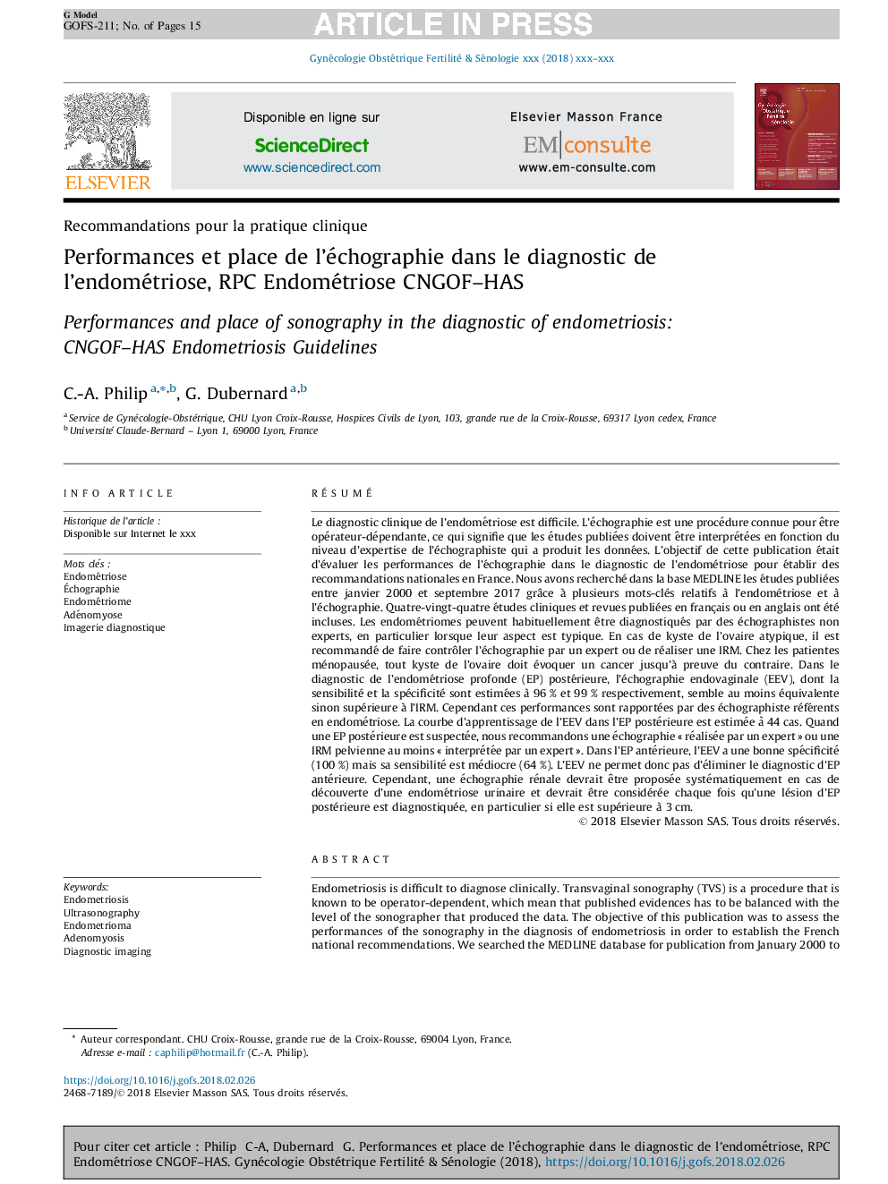
Endometriosis is difficult to diagnose clinically. Transvaginal sonography (TVS) is a procedure that is known to be operator-dependent, which mean that published evidences has to be balanced with the level of the sonographer that produced the data. The objective of this publication was to assess the performances of the sonography in the diagnosis of endometriosis in order to establish the French national recommendations. We searched the MEDLINE database for publication from January 2000 to September 2017 using keywords associated with endometriosis and sonography. Eighty-four trial and reviews published in English or French were included. Ovarian endometrioma can usually be diagnosed by a non-expert sonographer, especially when its aspect is typical. In case of an ovarian cyst with atypical presentation, it is recommended to control the sonography by a referent or to perform an MRI. In menopaused women, any ovarian cyst should be considered as a cancer until proven otherwise. In the diagnosis of posterior deep invasive endometriosis (DIE), TVS with sensitivity and specificity of 96 and 99% respectively, seems at least equivalent if not superior to MRI. However, these performances are related to expert sonographers. To reach sufficient efficiency in posterior DIE, the estimated learning curve for a sonographer is 44 cases. When posterior DIE is suspected, we recommend proposing a TVS “performed by an expert” or a MRI “at least interpreted by an expert”. In anterior DIE, TVS has a good specificity (100%), but its sensitivity is poor in the literature (64%). TVS is therefore not able to eliminate the diagnosis. However a renal ultrasound should be proposed each time a urinary endometriosis is confirmed, and should be considered whenever posterior DIE is diagnosed especially the lesion is superior to 3Â cm.
ناشر
Database: Elsevier - ScienceDirect (ساینس دایرکت)
Journal: Gynécologie Obstétrique Fertilité & Sénologie - Volume 46, Issue 3, March 2018, Pages 185-199
Journal: Gynécologie Obstétrique Fertilité & Sénologie - Volume 46, Issue 3, March 2018, Pages 185-199
نویسندگان
C.-A. Philip, G. Dubernard,