| Article ID | Journal | Published Year | Pages | File Type |
|---|---|---|---|---|
| 1229458 | Spectrochimica Acta Part A: Molecular and Biomolecular Spectroscopy | 2015 | 8 Pages |
•Sorafenib binds to DNA via minor groove binding and forms 1:1 complex with it.•The main interaction forces were van der Waals and hydrogen bonding interactions.•There was slight change of the secondary structure of DNA due to binding sorafenib.•The flexibility of sorafenib plays an important role in increasing the sorafenib–DNA stability.
The binding interaction of sorafenib with calf thymus DNA (ct-DNA) was studied using UV–vis absorption spectroscopy, fluorescence emission spectroscopy, circular dichroism (CD), viscosity measurement and molecular docking methods. The experimental results revealed that there was obvious binding interaction between sorafenib and ct-DNA. The binding constant (Kb) of sorafenib with ct-DNA was 5.6 × 10³ M–1 at 298 K. The enthalpy and entropy changes (ΔH0 and ΔS0) in the binding process of sorafenib with ct-DNA were –27.66 KJ mol–1 and –21.02 J mol–1 K–1, respectively, indicating that the main binding interaction forces were van der Waals force and hydrogen bonding. The docking results suggested that sorafenib preferred to bind on the minor groove of A-T rich DNA and the binding site of sorafenib was 4 base pairs long. The conformation change of sorafenib in the sorafenib–DNA complex was obviously observed and the change was close relation with the structure of DNA, implying that the flexibility of sorafenib molecule played an important role in the formation of the stable sorafenib–ct-DNA complex.
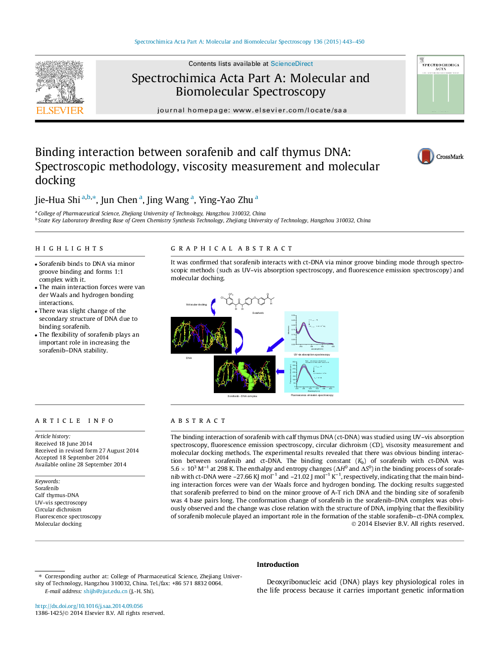
Graphical abstractIt was confirmed that sorafenib interacts with ct-DNA via minor groove binding mode through spectroscopic methods (such as UV–vis absorption spectroscopy, and fluorescence emission spectroscopy) and molecular doching.Figure optionsDownload full-size imageDownload as PowerPoint slide
