| Article ID | Journal | Published Year | Pages | File Type |
|---|---|---|---|---|
| 1760774 | Ultrasound in Medicine & Biology | 2011 | 14 Pages |
Abstract
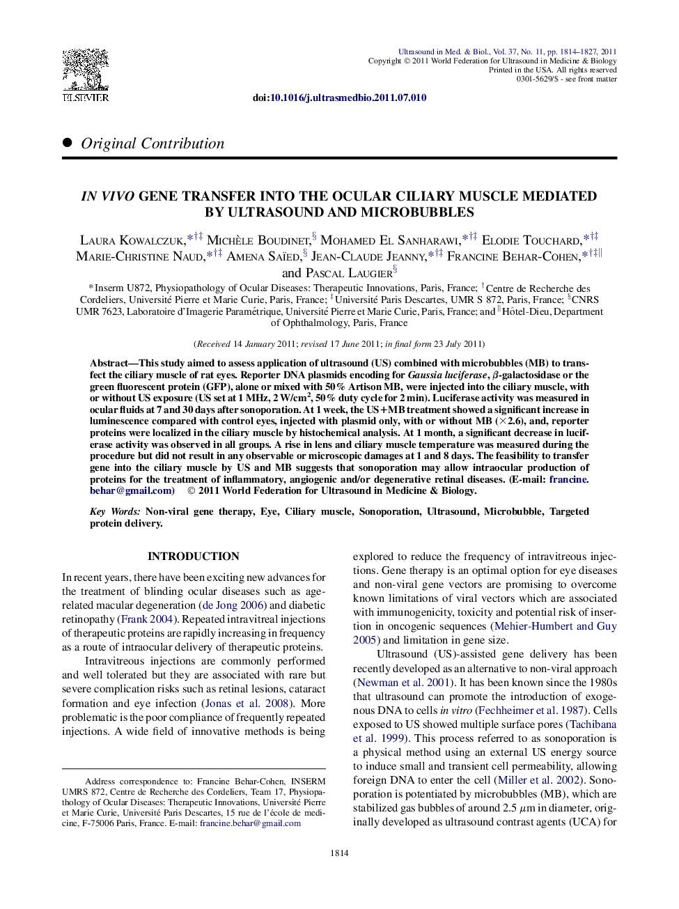
This study aimed to assess application of ultrasound (US) combined with microbubbles (MB) to transfect the ciliary muscle of rat eyes. Reporter DNA plasmids encoding for Gaussia luciferase, β-galactosidase or the green fluorescent protein (GFP), alone or mixed with 50% Artison MB, were injected into the ciliary muscle, with or without US exposure (US set at 1 MHz, 2 W/cm2, 50% duty cycle for 2 min). Luciferase activity was measured in ocular fluids at 7 and 30 days after sonoporation. At 1 week, the US+MB treatment showed a significant increase in luminescence compared with control eyes, injected with plasmid only, with or without MB (Ã2.6), and, reporter proteins were localized in the ciliary muscle by histochemical analysis. At 1 month, a significant decrease in luciferase activity was observed in all groups. A rise in lens and ciliary muscle temperature was measured during the procedure but did not result in any observable or microscopic damages at 1 and 8 days. The feasibility to transfer gene into the ciliary muscle by US and MB suggests that sonoporation may allow intraocular production of proteins for the treatment of inflammatory, angiogenic and/or degenerative retinal diseases.
Related Topics
Physical Sciences and Engineering
Physics and Astronomy
Acoustics and Ultrasonics
Authors
Laura Kowalczuk, Michèle Boudinet, Mohamed El Sanharawi, Elodie Touchard, Marie-Christine Naud, Amena Saïed, Jean-Claude Jeanny, Francine Behar-Cohen, Pascal Laugier,
