| Article ID | Journal | Published Year | Pages | File Type |
|---|---|---|---|---|
| 1762600 | Ultrasound in Medicine & Biology | 2007 | 7 Pages |
Abstract
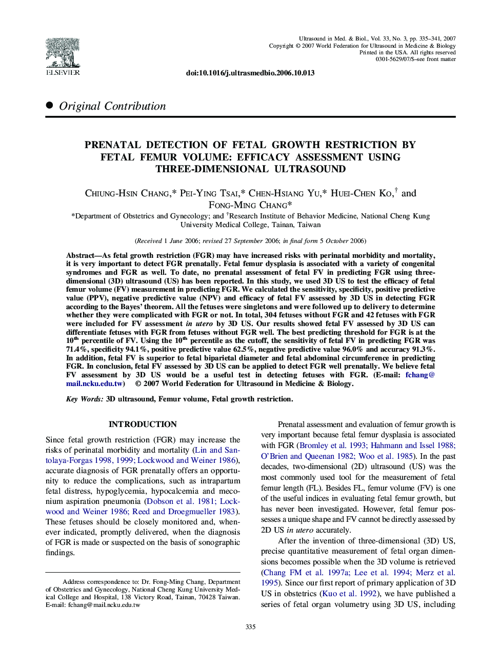
As fetal growth restriction (FGR) may have increased risks with perinatal morbidity and mortality, it is very important to detect FGR prenatally. Fetal femur dysplasia is associated with a variety of congenital syndromes and FGR as well. To date, no prenatal assessment of fetal FV in predicting FGR using three-dimensional (3D) ultrasound (US) has been reported. In this study, we used 3D US to test the efficacy of fetal femur volume (FV) measurement in predicting FGR. We calculated the sensitivity, specificity, positive predictive value (PPV), negative predictive value (NPV) and efficacy of fetal FV assessed by 3D US in detecting FGR according to the Bayes' theorem. All the fetuses were singletons and were followed up to delivery to determine whether they were complicated with FGR or not. In total, 304 fetuses without FGR and 42 fetuses with FGR were included for FV assessment in utero by 3D US. Our results showed fetal FV assessed by 3D US can differentiate fetuses with FGR from fetuses without FGR well. The best predicting threshold for FGR is at the 10th percentile of FV. Using the 10th percentile as the cutoff, the sensitivity of fetal FV in predicting FGR was 71.4%, specificity 94.1%, positive predictive value 62.5%, negative predictive value 96.0% and accuracy 91.3%. In addition, fetal FV is superior to fetal biparietal diameter and fetal abdominal circumference in predicting FGR. In conclusion, fetal FV assessed by 3D US can be applied to detect FGR well prenatally. We believe fetal FV assessment by 3D US would be a useful test in detecting fetuses with FGR. (E-mail: fchang@mail.ncku.edu.tw)
Related Topics
Physical Sciences and Engineering
Physics and Astronomy
Acoustics and Ultrasonics
Authors
Chiung-Hsin Chang, Pei-Ying Tsai, Chen-Hsiang Yu, Huei-Chen Ko, Fong-Ming Chang,
