| Article ID | Journal | Published Year | Pages | File Type |
|---|---|---|---|---|
| 2172013 | Cytotherapy | 2011 | 15 Pages |
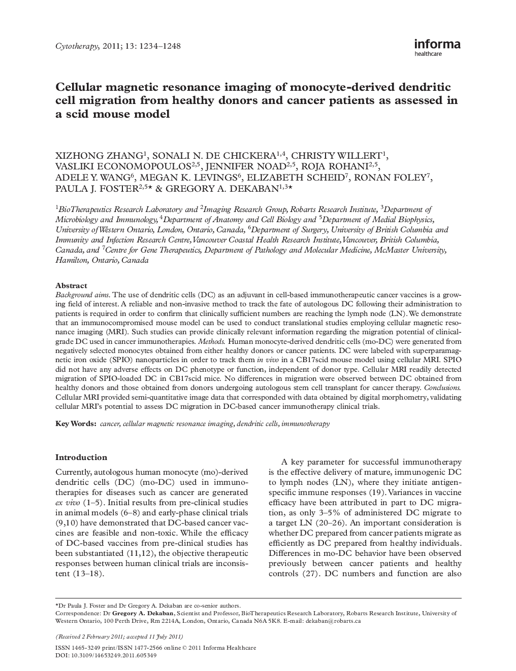
Background aimsThe use of dendritic cells (DC) as an adjuvant in cell-based immunotherapeutic cancer vaccines is a growing field of interest. A reliable and non-invasive method to track the fate of autologous DC following their administration to patients is required in order to confirm that clinically sufficient numbers are reaching the lymph node (LN). We demonstrate that an immunocompromised mouse model can be used to conduct translational studies employing cellular magnetic resonance imaging (MRI). Such studies can provide clinically relevant information regarding the migration potential of clinical-grade DC used in cancer immunotherapies.MethodsHuman monocyte-derived dendritic cells (mo-DC) were generated from negatively selected monocytes obtained from either healthy donors or cancer patients. DC were labeled with superparamagnetic iron oxide (SPIO) nanoparticles in order to track them in vivo in a CB17scid mouse model using cellular MRI. SPIO did not have any adverse effects on DC phenotype or function, independent of donor type. Cellular MRI readily detected migration of SPIO-loaded DC in CB17scid mice. No differences in migration were observed between DC obtained from healthy donors and those obtained from donors undergoing autologous stem cell transplant for cancer therapy.ConclusionsCellular MRI provided semi-quantitative image data that corresponded with data obtained by digital morphometry, validating cellular MRI's potential to assess DC migration in DC-based cancer immunotherapy clinical trials.
