| Article ID | Journal | Published Year | Pages | File Type |
|---|---|---|---|---|
| 2755259 | Clinical Lymphoma Myeloma and Leukemia | 2012 | 9 Pages |
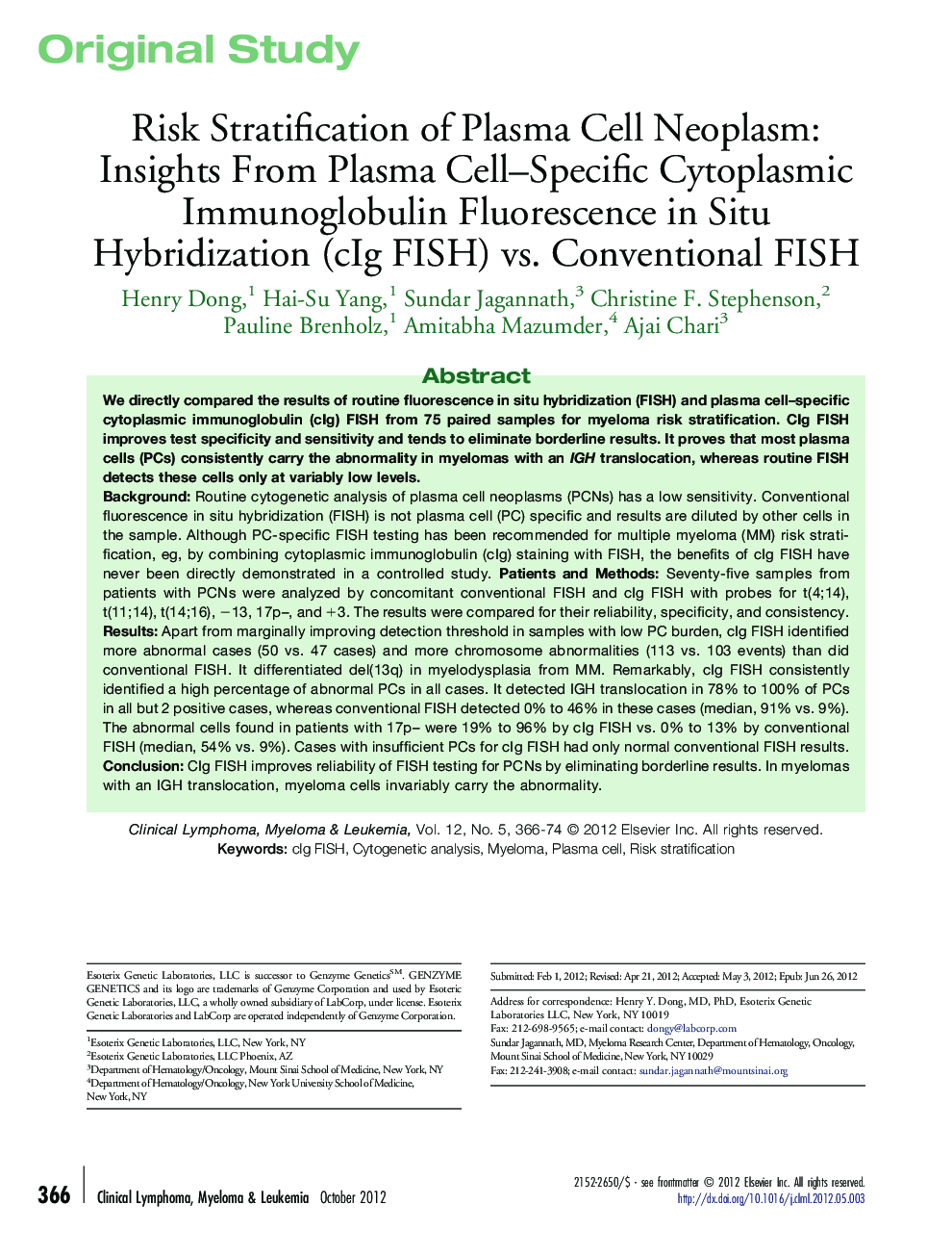
BackgroundRoutine cytogenetic analysis of plasma cell neoplasms (PCNs) has a low sensitivity. Conventional fluorescence in situ hybridization (FISH) is not plasma cell (PC) specific and results are diluted by other cells in the sample. Although PC-specific FISH testing has been recommended for multiple myeloma (MM) risk stratification, eg, by combining cytoplasmic immunoglobulin (cIg) staining with FISH, the benefits of cIg FISH have never been directly demonstrated in a controlled study.Patients and MethodsSeventy-five samples from patients with PCNs were analyzed by concomitant conventional FISH and cIg FISH with probes for t(4;14), t(11;14), t(14;16), −13, 17p–, and +3. The results were compared for their reliability, specificity, and consistency.ResultsApart from marginally improving detection threshold in samples with low PC burden, cIg FISH identified more abnormal cases (50 vs. 47 cases) and more chromosome abnormalities (113 vs. 103 events) than did conventional FISH. It differentiated del(13q) in myelodysplasia from MM. Remarkably, cIg FISH consistently identified a high percentage of abnormal PCs in all cases. It detected IGH translocation in 78% to 100% of PCs in all but 2 positive cases, whereas conventional FISH detected 0% to 46% in these cases (median, 91% vs. 9%). The abnormal cells found in patients with 17p– were 19% to 96% by cIg FISH vs. 0% to 13% by conventional FISH (median, 54% vs. 9%). Cases with insufficient PCs for cIg FISH had only normal conventional FISH results.ConclusionCIg FISH improves reliability of FISH testing for PCNs by eliminating borderline results. In myelomas with an IGH translocation, myeloma cells invariably carry the abnormality.
