| Article ID | Journal | Published Year | Pages | File Type |
|---|---|---|---|---|
| 2776904 | Journal of Oral Biosciences | 2011 | 12 Pages |
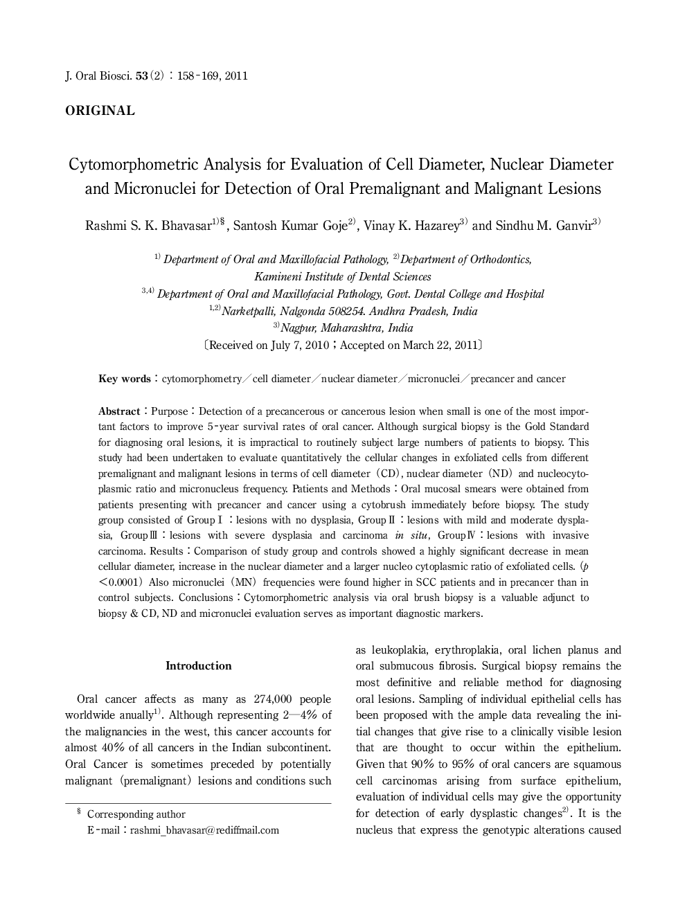
Purpose: Detection of a precancerous or cancerous lesion when small is one of the most important factors to improve 5-year survival rates of oral cancer. Although surgical biopsy is the Gold Standard for diagnosing oral lesions, it is impractical to routinely subject large numbers of patients to biopsy. This study had been undertaken to evaluate quantitatively the cellular changes in exfoliated cells from different premalignant and malignant lesions in terms of cell diameter (CD), nuclear diameter (ND) and nucleocytoplasmic ratio and micronucleus frequency. Patients and Methods: Oral mucosal smears were obtained from patients presenting with precancer and cancer using a cytobrush immediately before biopsy. The study group consisted of Group I: lesions with no dysplasia, Group It: lesions with mild and moderate dysplasia, Group Ul: lesions with severe dysplasia and carcinoma in situ, Group IV: lesions with invasive carcinoma. Results: Comparison of study group and controls showed a highly significant decrease in mean cellular diameter, increase in the nuclear diameter and a larger nucleo cytoplasmic ratio of exfoliated cells. (p <0.0001) Also micronuclei (MN) frequencies were found higher in SCC patients and in precancer than in control subjects. Conclusions: Cytomorphometric analysis via oral brush biopsy is a valuable adjunct to biopsy & CD, ND and micronuclei evaluation serves as important diagnostic markers.
