| Article ID | Journal | Published Year | Pages | File Type |
|---|---|---|---|---|
| 2906015 | Chest | 2006 | 6 Pages |
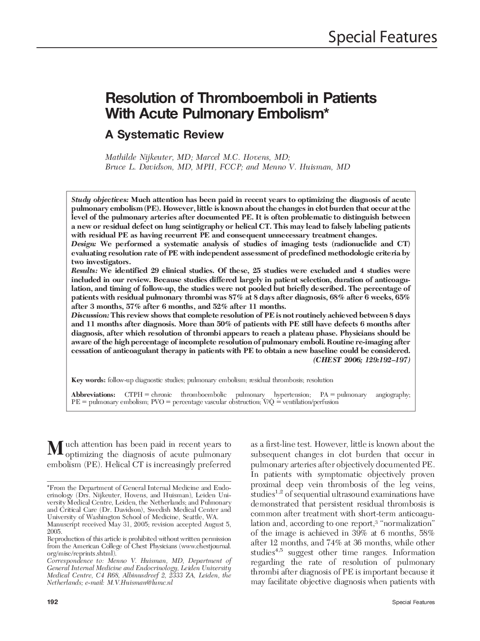
Study objectivesMuch attention has been paid in recent years to optimizing the diagnosis of acute pulmonary embolism (PE). However, little is known about the changes in clot burden that occur at the level of the pulmonary arteries after documented PE. It is often problematic to distinguish between a new or residual defect on lung scintigraphy or helical CT. This may lead to falsely labeling patients with residual PE as having recurrent PE and consequent unnecessary treatment changes.DesignWe performed a systematic analysis of studies of imaging tests (radionuclide and CT) evaluating resolution rate of PE with independent assessment of predefined methodologic criteria by two investigators.ResultsWe identified 29 clinical studies. Of these, 25 studies were excluded and 4 studies were included in our review. Because studies differed largely in patient selection, duration of anticoagulation, and timing of follow-up, the studies were not pooled but briefly described. The percentage of patients with residual pulmonary thrombi was 87% at 8 days after diagnosis, 68% after 6 weeks, 65% after 3 months, 57% after 6 months, and 52% after 11 months.DiscussionThis review shows that complete resolution of PE is not routinely achieved between 8 days and 11 months after diagnosis. More than 50% of patients with PE still have defects 6 months after diagnosis, after which resolution of thrombi appears to reach a plateau phase. Physicians should be aware of the high percentage of incomplete resolution of pulmonary emboli. Routine re-imaging after cessation of anticoagulant therapy in patients with PE to obtain a new baseline could be considered.
