| Article ID | Journal | Published Year | Pages | File Type |
|---|---|---|---|---|
| 2939251 | JACC: Cardiovascular Imaging | 2009 | 10 Pages |
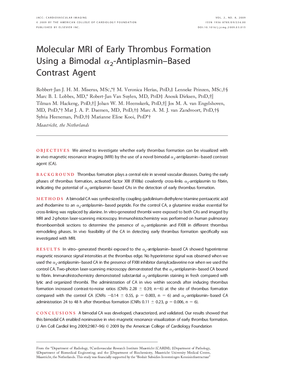
ObjectivesWe aimed to investigate whether early thrombus formation can be visualized with in vivo magnetic resonance imaging (MRI) by the use of a novel bimodal α2-antiplasmin–based contrast agent (CA).BackgroundThrombus formation plays a central role in several vascular diseases. During the early phases of thrombus formation, activated factor XIII (FXIIIa) covalently cross-links α2-antiplasmin to fibrin, indicating the potential of α2-antiplasmin–based CAs in the detection of early thrombus formation.MethodsA bimodal CA was synthesized by coupling gadolinium-diethylene triamine pentaacetic acid and rhodamine to an α2-antiplasmin–based peptide. For the control CA, a glutamine residue essential for cross-linking was replaced by alanine. In vitro-generated thrombi were exposed to both CAs and imaged by MRI and 2-photon laser-scanning microscopy. Immunohistochemistry was performed on human pulmonary thromboemboli sections to determine the presence of α2-antiplasmin and FXIII in different thrombus remodeling phases. In vivo feasibility of the CA in detecting early thrombus formation specifically was investigated with MRI.ResultsIn vitro–generated thrombi exposed to the α2-antiplasmin–based CA showed hyperintense magnetic resonance signal intensities at the thrombus edge. No hyperintense signal was observed when we used the α2-antiplasmin–based CA in the presence of FXIII inhibitor dansylcadaverine nor when we used the control CA. Two-photon laser-scanning microscopy demonstrated that the α2-antiplasmin–based CA bound to fibrin. Immunohistochemistry demonstrated substantial α2-antiplasmin staining in fresh compared with lytic and organized thrombi. The administration of CA in vivo within seconds after inducing thrombus formation increased contrast-to-noise ratios (CNRs 2.28 ± 0.39, n=6) at the site of thrombus formation compared with the control CA (CNRs −0.14 ± 0.55, p = 0.003, n = 6) and α2-antiplasmin–based CA administration 24 to 48 h after thrombus formation (CNRs 0.11 ± 0.23, p = 0.006, n = 6).ConclusionsA bimodal CA was developed, characterized, and validated. Our results showed that this bimodal CA enabled noninvasive in vivo magnetic resonance visualization of early thrombus formation.
