| Article ID | Journal | Published Year | Pages | File Type |
|---|---|---|---|---|
| 3062978 | Journal of Clinical Neuroscience | 2007 | 4 Pages |
Abstract
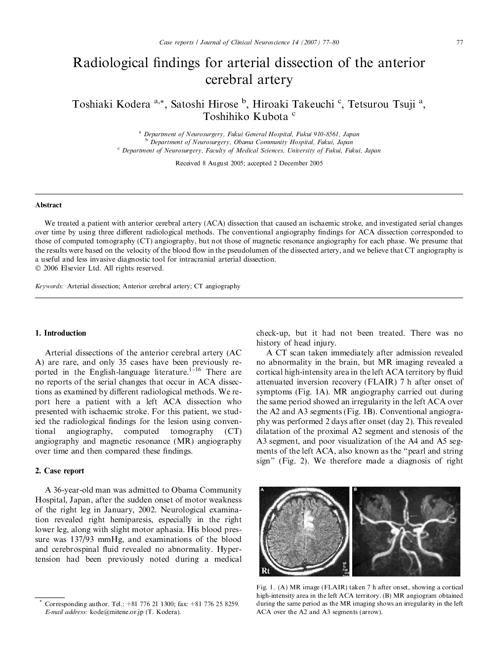
We treated a patient with anterior cerebral artery (ACA) dissection that caused an ischaemic stroke, and investigated serial changes over time by using three different radiological methods. The conventional angiography findings for ACA dissection corresponded to those of computed tomography (CT) angiography, but not those of magnetic resonance angiography for each phase. We presume that the results were based on the velocity of the blood flow in the pseudolumen of the dissected artery, and we believe that CT angiography is a useful and less invasive diagnostic tool for intracranial arterial dissection.
Related Topics
Life Sciences
Neuroscience
Neurology
Authors
Toshiaki Kodera, Satoshi Hirose, Hiroaki Takeuchi, Tetsurou Tsuji, Toshihiko Kubota,
