| Article ID | Journal | Published Year | Pages | File Type |
|---|---|---|---|---|
| 3071832 | Neurocirugía | 2010 | 6 Pages |
Abstract
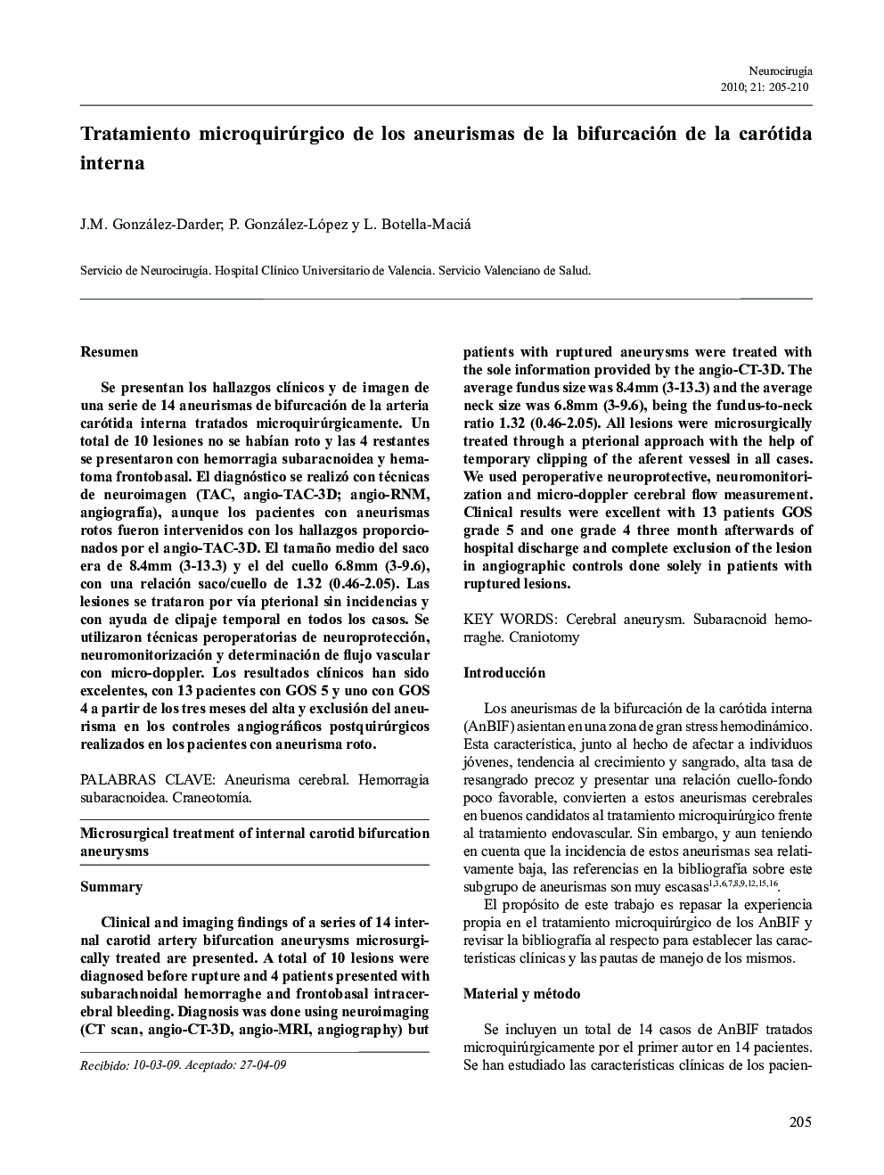
Clinical and imaging findings of a series of 14 internal carotid artery bifurcation aneurysms microsurgically treated are presented. A total of 10 lesions were diagnosed before rupture and 4 patients presented with subarachnoidal hemorraghe and frontobasal intracerebral bleeding. Diagnosis was done using neuroimaging (CT scan, angio-CT-3D, angio-MRI, angiography) but patients with ruptured aneurysms were treated with the sole information provided by the angio-CT-3D. The average fundus size was 8.4Â mm (3-13.3) and the average neck size was 6.8Â mm (3-9.6), being the fundus-to-neck ratio 1.32 (0.46-2.05). All lesions were microsurgically treated through a pterional approach with the help of temporary clipping of the aferent vessesl in all cases. We used peroperative neuroprotective, neuromonitorization and micro-doppler cerebral flow measurement. Clinical results were excellent with 13 patients GOS grade 5 and one grade 4 three month afterwards of hospital discharge and complete exclusion of the lesion in angiographic controls done solely in patients with ruptured lesions.
Related Topics
Life Sciences
Neuroscience
Neurology
Authors
J.M. (Dr), P. González-López, L. Botella-Maciá,
