| Article ID | Journal | Published Year | Pages | File Type |
|---|---|---|---|---|
| 3093681 | Surgical Neurology | 2007 | 5 Pages |
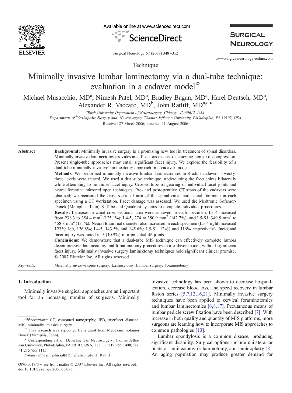
BackgroundMinimally invasive surgery is a promising new tool in treatment of spinal disorders. Minimally invasive laminectomy provides an efficacious means of achieving lumbar decompression. Present single-tube approaches may entail significant facet injury. We explore the feasibility of a dual-tube minimally invasive laminectomy approach in a cadaver model.MethodsWe performed minimally invasive lumbar laminectomies in 8 adult cadavers. Twenty-three levels were treated. We used a dual-tube technique, undercutting the facet joints bilaterally while attempting to minimize facet injury. Crossed-tube rongeuring of individual facet joints and neural foramina mirrored open techniques. Pre- and postoperative CT scans of the cadavers were obtained; we measured the cross-sectional area of the spinal canal and neural foramina in each specimen using a CT workstation. Facet damage was assessed. We used the Medtronic Sofamor-Danek (Memphis, Tenn) X-Tube and Quadrant systems to complete individual procedures.ResultsIncreases in canal cross-sectional area were achieved in each specimen: L3-4 increased from 238.3 to 354.4 mm2 (125.1%); L4-5, 274 to 390.9 mm2 (142.7%); and L5-S1, 349.9 mm2 to 458.8 mm2 (131%). Neural foraminal diameter also increased in each specimen (L3-4 right increased 123%; left, 136.8%; L4-5, 143.5% and 145.6%; L5-S1, 124% and 116% respectively). Incidental facet injury was noted in 5 (10.9%) of a potential 46 joints.ConclusionsWe demonstrate that a dual-tube MIS technique can effectively complete lumbar decompressive laminectomy and foraminotomy procedures in a cadaver model, without significant facet injury. Minimally invasive surgery laminectomy techniques hold significant clinical promise.
