| Article ID | Journal | Published Year | Pages | File Type |
|---|---|---|---|---|
| 3097510 | World Neurosurgery | 2010 | 4 Pages |
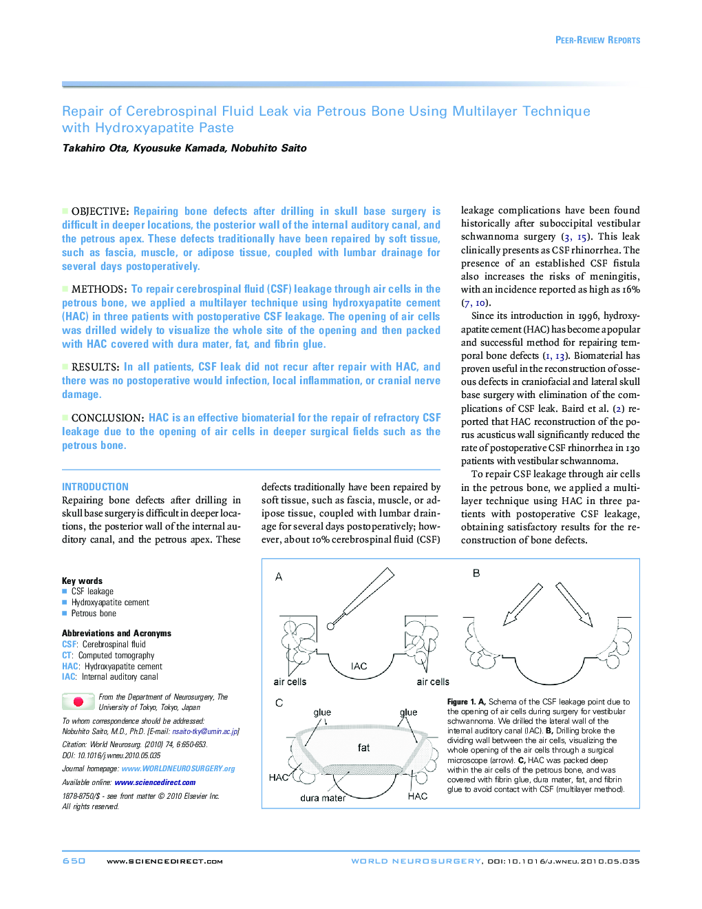
ObjectiveRepairing bone defects after drilling in skull base surgery is difficult in deeper locations, the posterior wall of the internal auditory canal, and the petrous apex. These defects traditionally have been repaired by soft tissue, such as fascia, muscle, or adipose tissue, coupled with lumbar drainage for several days postoperatively.MethodsTo repair cerebrospinal fluid (CSF) leakage through air cells in the petrous bone, we applied a multilayer technique using hydroxyapatite cement (HAC) in three patients with postoperative CSF leakage. The opening of air cells was drilled widely to visualize the whole site of the opening and then packed with HAC covered with dura mater, fat, and fibrin glue.ResultsIn all patients, CSF leak did not recur after repair with HAC, and there was no postoperative would infection, local inflammation, or cranial nerve damage.ConclusionHAC is an effective biomaterial for the repair of refractory CSF leakage due to the opening of air cells in deeper surgical fields such as the petrous bone.
