| Article ID | Journal | Published Year | Pages | File Type |
|---|---|---|---|---|
| 3166318 | Oral Surgery, Oral Medicine, Oral Pathology and Oral Radiology | 2016 | 9 Pages |
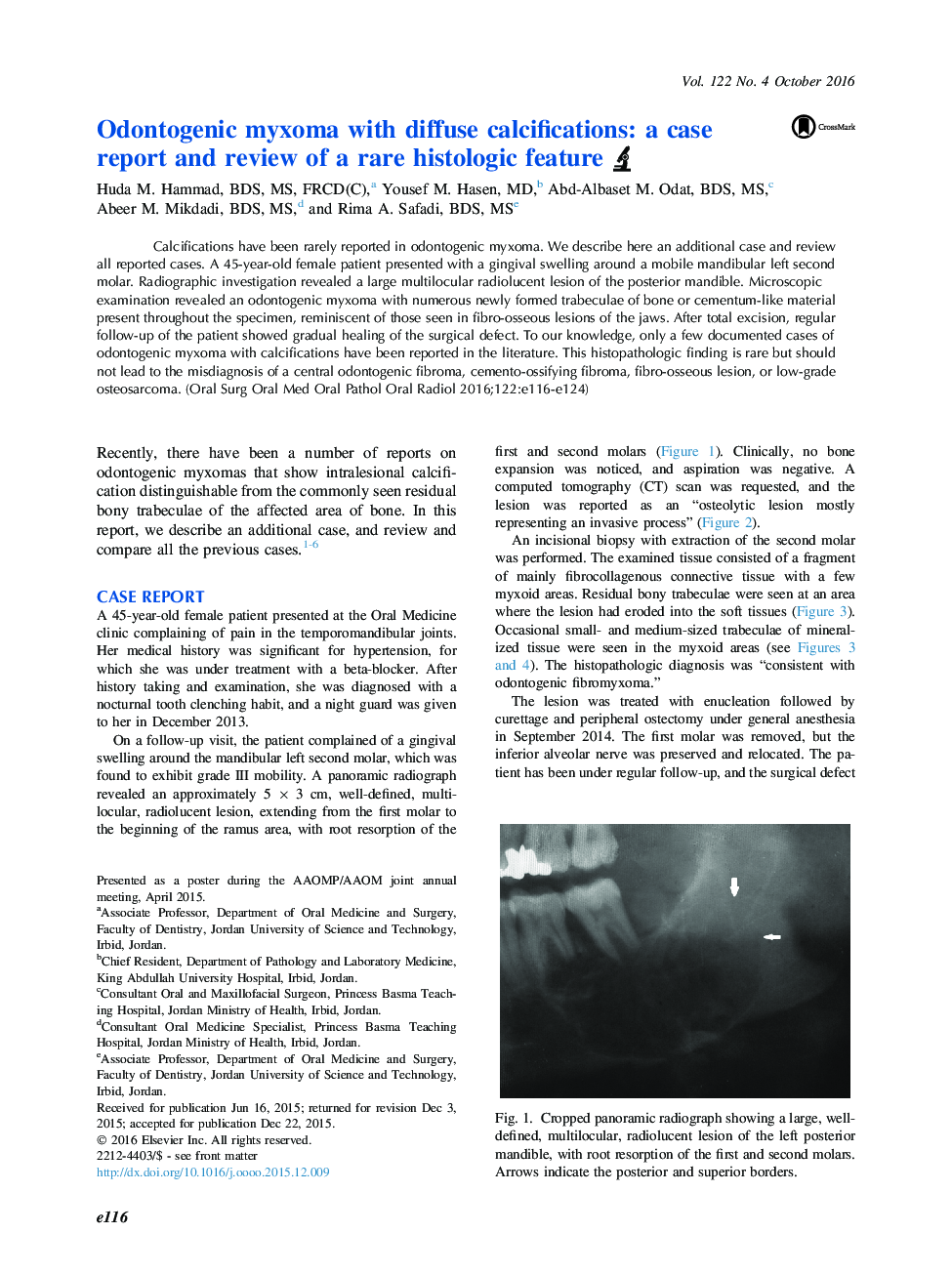
Calcifications have been rarely reported in odontogenic myxoma. We describe here an additional case and review all reported cases. A 45-year-old female patient presented with a gingival swelling around a mobile mandibular left second molar. Radiographic investigation revealed a large multilocular radiolucent lesion of the posterior mandible. Microscopic examination revealed an odontogenic myxoma with numerous newly formed trabeculae of bone or cementum-like material present throughout the specimen, reminiscent of those seen in fibro-osseous lesions of the jaws. After total excision, regular follow-up of the patient showed gradual healing of the surgical defect. To our knowledge, only a few documented cases of odontogenic myxoma with calcifications have been reported in the literature. This histopathologic finding is rare but should not lead to the misdiagnosis of a central odontogenic fibroma, cemento-ossifying fibroma, fibro-osseous lesion, or low-grade osteosarcoma.
