| Article ID | Journal | Published Year | Pages | File Type |
|---|---|---|---|---|
| 3204763 | Journal of the American Academy of Dermatology | 2015 | 7 Pages |
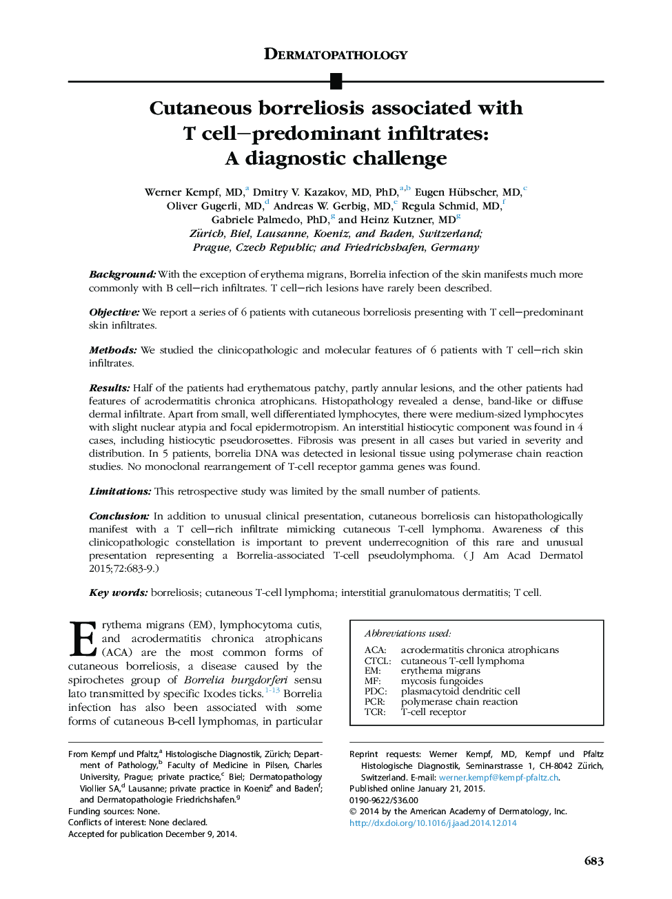
BackgroundWith the exception of erythema migrans, Borrelia infection of the skin manifests much more commonly with B cell–rich infiltrates. T cell–rich lesions have rarely been described.ObjectiveWe report a series of 6 patients with cutaneous borreliosis presenting with T cell–predominant skin infiltrates.MethodsWe studied the clinicopathologic and molecular features of 6 patients with T cell–rich skin infiltrates.ResultsHalf of the patients had erythematous patchy, partly annular lesions, and the other patients had features of acrodermatitis chronica atrophicans. Histopathology revealed a dense, band-like or diffuse dermal infiltrate. Apart from small, well differentiated lymphocytes, there were medium-sized lymphocytes with slight nuclear atypia and focal epidermotropism. An interstitial histiocytic component was found in 4 cases, including histiocytic pseudorosettes. Fibrosis was present in all cases but varied in severity and distribution. In 5 patients, borrelia DNA was detected in lesional tissue using polymerase chain reaction studies. No monoclonal rearrangement of T-cell receptor gamma genes was found.LimitationsThis retrospective study was limited by the small number of patients.ConclusionIn addition to unusual clinical presentation, cutaneous borreliosis can histopathologically manifest with a T cell–rich infiltrate mimicking cutaneous T-cell lymphoma. Awareness of this clinicopathologic constellation is important to prevent underrecognition of this rare and unusual presentation representing a Borrelia-associated T-cell pseudolymphoma.
