| Article ID | Journal | Published Year | Pages | File Type |
|---|---|---|---|---|
| 3317737 | Pancreatology | 2011 | 9 Pages |
Abstract
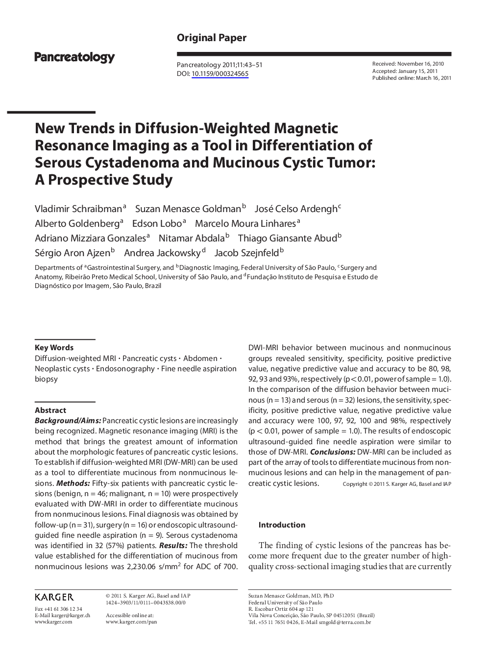
Background/Aims: Pancreatic cystic lesions are increasingly being recognized. Magnetic resonance imaging (MRI) is the method that brings the greatest amount of information about the morphologic features of pancreatic cystic lesions. To establish if diffusion-weighted MRI (DW-MRI) can be used as a tool to differentiate mucinous from nonmucinous lesions. Methods: Fifty-six patients with pancreatic cystic lesions (benign, n = 46; malignant, n = 10) were prospectively evaluated with DW-MRI in order to differentiate mucinous from nonmucinous lesions. Final diagnosis was obtained by follow-up (n = 31), surgery (n = 16) or endoscopic ultrasound-guided fine needle aspiration (n = 9). Serous cystadenoma was identified in 32 (57%) patients. Results: The threshold value established for the differentiation of mucinous from nonmucinous lesions was 2,230.06 s/mm2 for ADC of 700. DWI-MRI behavior between mucinous and nonmucinous groups revealed sensitivity, specificity, positive predictive value, negative predictive value and accuracy to be 80, 98, 92,93 and 93%, respectively (p < 0.01, power of sample = 1.0). In the comparison of the diffusion behavior between mucinous (n = 13) and serous (n = 32) lesions, the sensitivity, specificity, positive predictive value, negative predictive value and accuracy were 100, 97, 92, 100 and 98%, respectively (p < 0.01, power of sample = 1.0). The results of endoscopic ultrasound-guided fine needle aspiration were similar to those of DW-MRI. Conclusions: DW-MRI can be included as part of the array of tools to differentiate mucinous from nonmucinous lesions and can help in the management of pancreatic cystic lesions.
Related Topics
Health Sciences
Medicine and Dentistry
Gastroenterology
Authors
Vladimir Schraibman, Suzan Menasce Goldman, José Celso Ardengh, Alberto Goldenberg, Edson Lobo, Marcelo Moura Linhares, Adriano Mizziara Gonzales, Nitamar Abdala, Thiago Giansante Abud, Sérgio Aron Ajzen, Andrea Jackowsky, Jacob Szejnfeld,
