| Article ID | Journal | Published Year | Pages | File Type |
|---|---|---|---|---|
| 3337426 | Hepatobiliary & Pancreatic Diseases International | 2014 | 8 Pages |
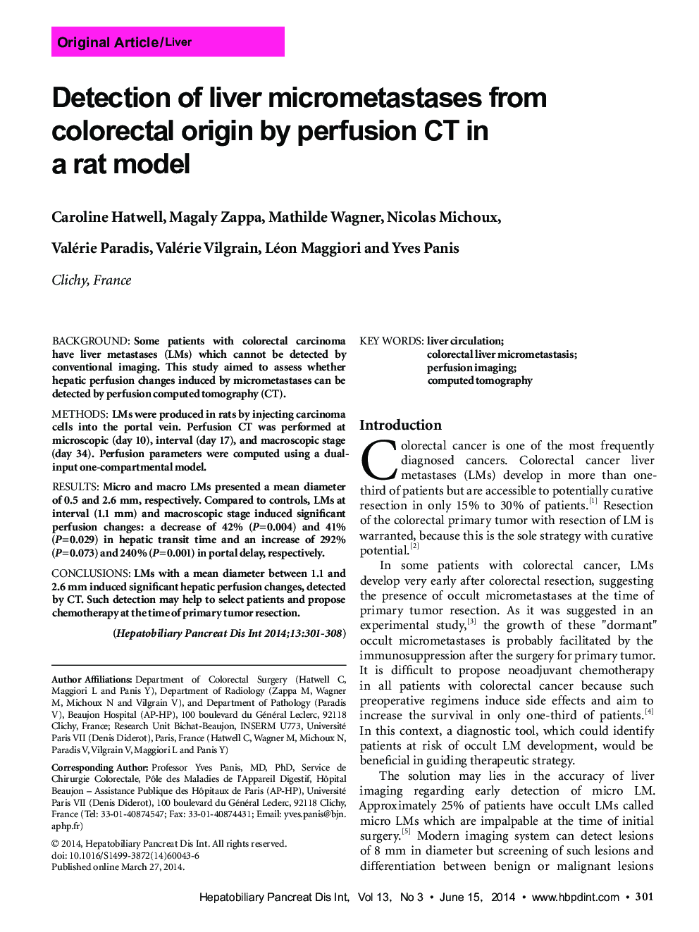
BackgroundSome patients with colorectal carcinoma have liver metastases (LMs) which cannot be detected by conventional imaging. This study aimed to assess whether hepatic perfusion changes induced by micrometastases can be detected by perfusion computed tomography (CT).MethodsLMs were produced in rats by injecting carcinoma cells into the portal vein. Perfusion CT was performed at microscopic (day 10), interval (day 17), and macroscopic stage (day 34). Perfusion parameters were computed using a dualinput one-compartmental model.ResultsMicro and macro LMs presented a mean diameter of 0.5 and 2.6 mm, respectively. Compared to controls, LMs at interval (1.1 mm) and macroscopic stage induced significant perfusion changes: a decrease of 42% (P=0.004) and 41% (P=0.029) in hepatic transit time and an increase of 292% (P=0.073) and 240% (P=0.001) in portal delay, respectively.ConclusionsLMs with a mean diameter between 1.1 and 2.6 mm induced significant hepatic perfusion changes, detected by CT. Such detection may help to select patients and propose chemotherapy at the time of primary tumor resection.
