| Article ID | Journal | Published Year | Pages | File Type |
|---|---|---|---|---|
| 3459657 | Chinese Medical Sciences Journal | 2008 | 5 Pages |
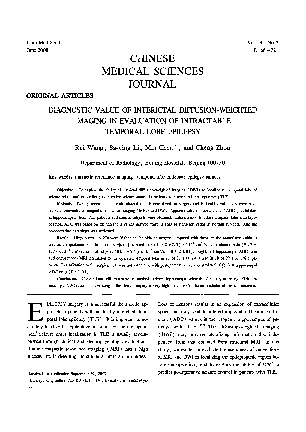
ObjectiveTo explore the ability of interictal diffusion-weighted imaging (DWI) to localize the temporal lobe of seizure origin and to predict postoperative seizure control in patients with temporal lobe epilepsy (TLE).MethodsTwenty-seven patients with intractable TLE considered for surgery and 19 healthy volunteers were studied with conventional magnetic resonance imaging (MRI) and DWI. Apparent diffusion coefficients (ADCs) of bilateral hippocampi in both TLE patients and control subjects were obtained. Lateralization to either temporal lobe with hipp-ocampal ADC was based on the threshold values derived from ± 1SD of right/left ratios in normal subjects. And the postoperative pathology was reviewed.ResultsHippocampal ADCs were higher on the side of surgery compared with those on the contralateral side as well as the ipsilateral side in control subjects [resected side (109.8 ± 7.3) × 10−5 cm2/s, contralateral side (91.7 ± 4.7) × 10−5 cm2/s, control subjects (81.6 ± 5.2) × 10−5 cm2/s, all P < 0.01]. Right/left hippocampal ADC ratio and conventional MRI lateralized to the operated temporal lobe in 21 of 27 (77.8%) and in 18 of 27 (66.7%) patients. Lateralization to the surgical side was not associated with postoperative seizure control with right/left hippocampal ADC ratio (P >0.05).ConclusionConventional MRI is a sensitive method to detect hippocampal sclerosis. Accuracy of the right/left hippocampal ADC ratio for lateralizing to the side of surgery is very high, but it isn't a better predictor of surgical outcome.
