| Article ID | Journal | Published Year | Pages | File Type |
|---|---|---|---|---|
| 3859810 | The Journal of Urology | 2015 | 7 Pages |
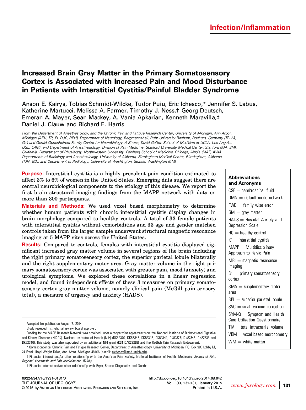
PurposeInterstitial cystitis is a highly prevalent pain condition estimated to affect 3% to 6% of women in the United States. Emerging data suggest there are central neurobiological components to the etiology of this disease. We report the first brain structural imaging findings from the MAPP network with data on more than 300 participants.Materials and MethodsWe used voxel based morphometry to determine whether human patients with chronic interstitial cystitis display changes in brain morphology compared to healthy controls. A total of 33 female patients with interstitial cystitis without comorbidities and 33 age and gender matched controls taken from the larger sample underwent structural magnetic resonance imaging at 5 MAPP sites across the United States.ResultsCompared to controls, females with interstitial cystitis displayed significant increased gray matter volume in several regions of the brain including the right primary somatosensory cortex, the superior parietal lobule bilaterally and the right supplementary motor area. Gray matter volume in the right primary somatosensory cortex was associated with greater pain, mood (anxiety) and urological symptoms. We explored these correlations in a linear regression model, and found independent effects of these 3 measures on primary somatosensory cortex gray matter volume, namely clinical pain (McGill pain sensory total), a measure of urgency and anxiety (HADS).ConclusionsThese data support the notion that changes in somatosensory gray matter may have an important role in pain sensitivity as well as affective and sensory aspects of interstitial cystitis. Further studies are needed to confirm the generalizability of these findings to other pain conditions.
