| Article ID | Journal | Published Year | Pages | File Type |
|---|---|---|---|---|
| 3862384 | The Journal of Urology | 2015 | 7 Pages |
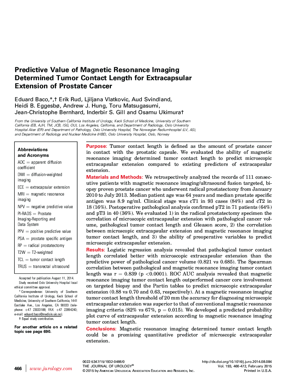
PurposeTumor contact length is defined as the amount of prostate cancer in contact with the prostatic capsule. We evaluated the ability of magnetic resonance imaging determined tumor contact length to predict microscopic extracapsular extension compared to existing predictors of extracapsular extension.Materials and MethodsWe retrospectively analyzed the records of 111 consecutive patients with magnetic resonance imaging/ultrasound fusion targeted, biopsy proven prostate cancer who underwent radical prostatectomy from January 2010 to July 2013. Median patient age was 64 years and median prostate specific antigen was 8.9 ng/ml. Clinical stage was cT1 in 93 cases (84%) and cT2 in 18 (16%). Postoperative pathological analysis confirmed pT2 in 71 patients (64%) and pT3 in 40 (36%). We evaluated 1) in the radical prostatectomy specimen the correlation of microscopic extracapsular extension with pathological cancer volume, pathological tumor contact length and Gleason score, 2) the correlation between microscopic extracapsular extension and magnetic resonance imaging tumor contact length, and 3) the ability of preoperative variables to predict microscopic extracapsular extension.ResultsLogistic regression analysis revealed that pathological tumor contact length correlated better with microscopic extracapsular extension than the predictive power of pathological cancer volume (0.821 vs 0.685). The Spearman correlation between pathological and magnetic resonance imaging tumor contact length was r = 0.839 (p <0.0001). ROC AUC analysis revealed that magnetic resonance imaging tumor contact length outperformed cancer core involvement on targeted biopsy and the Partin tables to predict microscopic extracapsular extension (0.88 vs 0.70 and 0.63, respectively). At a magnetic resonance imaging tumor contact length threshold of 20 mm the accuracy for diagnosing microscopic extracapsular extension was superior to that of conventional magnetic resonance imaging criteria (82% vs 67%, p = 0.015). We developed a predicted probability plot curve of extracapsular extension according to magnetic resonance imaging tumor contact length.ConclusionsMagnetic resonance imaging determined tumor contact length could be a promising quantitative predictor of microscopic extracapsular extension.
