| Article ID | Journal | Published Year | Pages | File Type |
|---|---|---|---|---|
| 3900041 | Urology | 2013 | 5 Pages |
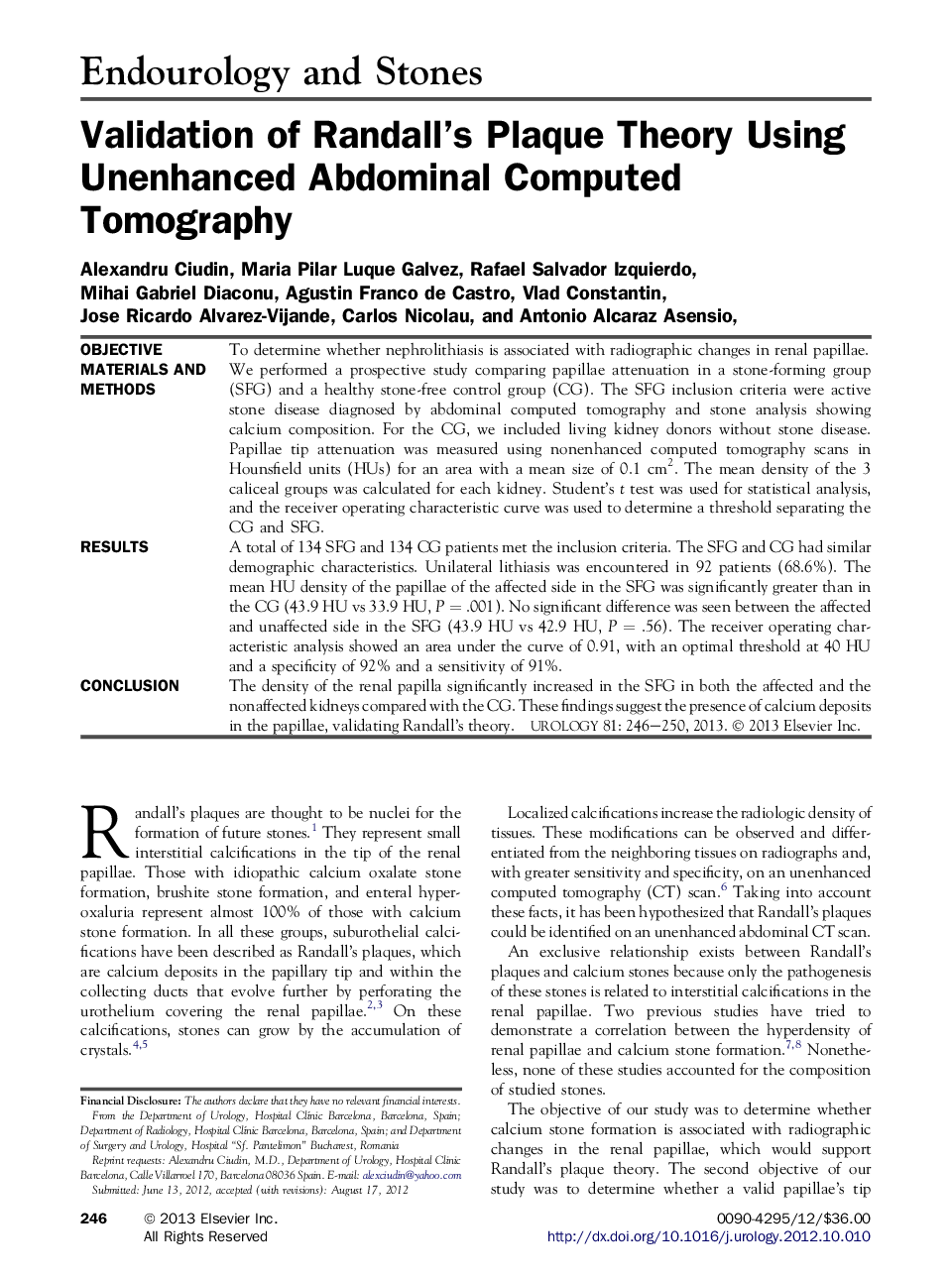
ObjectiveTo determine whether nephrolithiasis is associated with radiographic changes in renal papillae.Materials and MethodsWe performed a prospective study comparing papillae attenuation in a stone-forming group (SFG) and a healthy stone-free control group (CG). The SFG inclusion criteria were active stone disease diagnosed by abdominal computed tomography and stone analysis showing calcium composition. For the CG, we included living kidney donors without stone disease. Papillae tip attenuation was measured using nonenhanced computed tomography scans in Hounsfield units (HUs) for an area with a mean size of 0.1 cm2. The mean density of the 3 caliceal groups was calculated for each kidney. Student's t test was used for statistical analysis, and the receiver operating characteristic curve was used to determine a threshold separating the CG and SFG.ResultsA total of 134 SFG and 134 CG patients met the inclusion criteria. The SFG and CG had similar demographic characteristics. Unilateral lithiasis was encountered in 92 patients (68.6%). The mean HU density of the papillae of the affected side in the SFG was significantly greater than in the CG (43.9 HU vs 33.9 HU, P = .001). No significant difference was seen between the affected and unaffected side in the SFG (43.9 HU vs 42.9 HU, P = .56). The receiver operating characteristic analysis showed an area under the curve of 0.91, with an optimal threshold at 40 HU and a specificity of 92% and a sensitivity of 91%.ConclusionThe density of the renal papilla significantly increased in the SFG in both the affected and the nonaffected kidneys compared with the CG. These findings suggest the presence of calcium deposits in the papillae, validating Randall's theory.
