| Article ID | Journal | Published Year | Pages | File Type |
|---|---|---|---|---|
| 3975853 | Taiwanese Journal of Obstetrics and Gynecology | 2010 | 5 Pages |
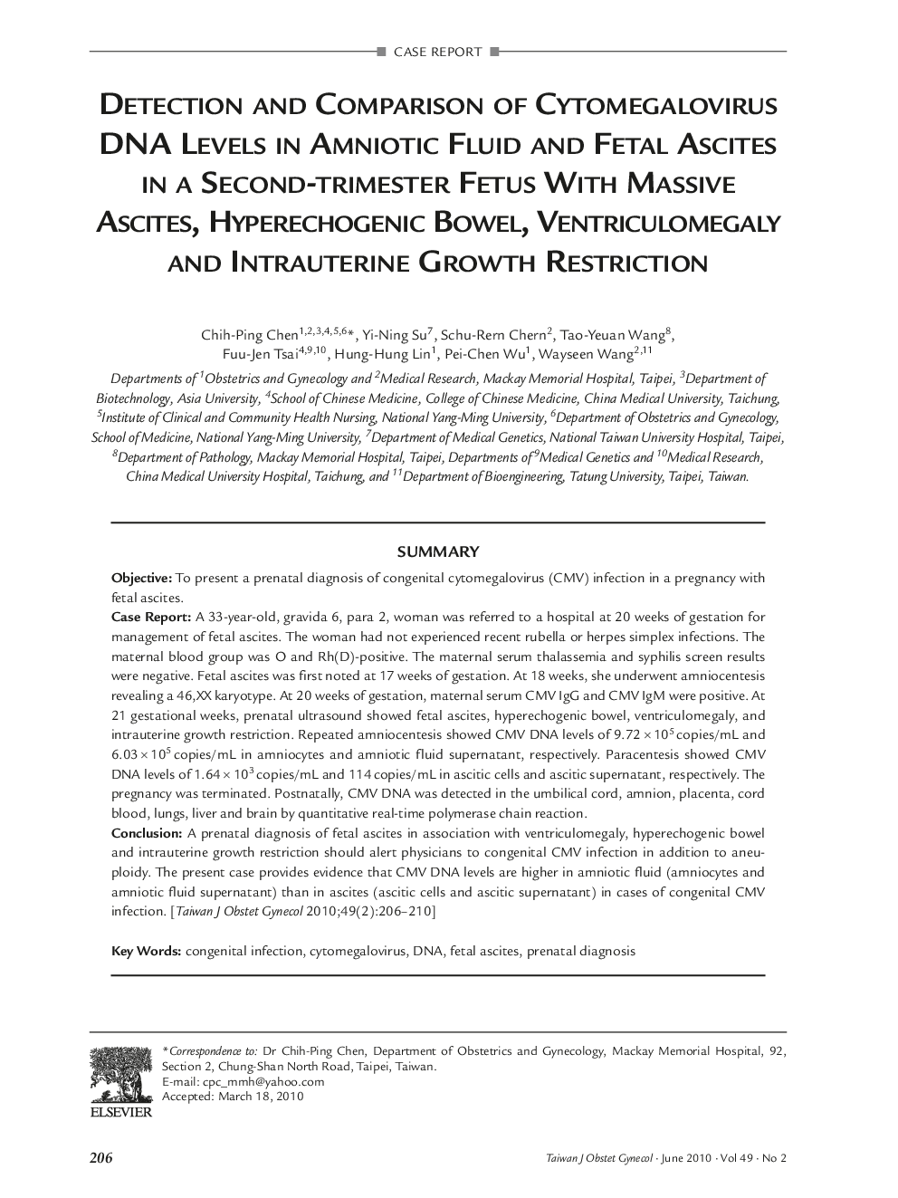
SummaryObjectiveTo present a prenatal diagnosis of congenital cytomegalovirus (CMV) infection in a pregnancy with fetal ascites.Case ReportA 33-year-old, gravida 6, para 2, woman was referred to a hospital at 20 weeks of gestation for management of fetal ascites. The woman had not experienced recent rubella or herpes simplex infections. The maternal blood group was O and Rh(D)-positive. The maternal serum thalassemia and syphilis screen results were negative. Fetal ascites was first noted at 17 weeks of gestation. At 18 weeks, she underwent amniocentesis revealing a 46,XX karyotype. At 20 weeks of gestation, maternal serum CMV IgG and CMV IgM were positive. At 21 gestational weeks, prenatal ultrasound showed fetal ascites, hyperechogenic bowel, ventriculomegaly, and intrauterine growth restriction. Repeated amniocentesis showed CMV DNA levels of 9.72 × 10⁵ copies/mL and 6.03 × 10⁵ copies/mL in amniocytes and amniotic fluid supernatant, respectively. Paracentesis showed CMV DNA levels of 1.64 × 10³ copies/mL and 114 copies/mL in ascitic cells and ascitic supernatant, respectively. The pregnancy was terminated. Postnatally, CMV DNA was detected in the umbilical cord, amnion, placenta, cord blood, lungs, liver and brain by quantitative real-time polymerase chain reaction.ConclusionA prenatal diagnosis of fetal ascites in association with ventriculomegaly, hyperechogenic bowel and intrauterine growth restriction should alert physicians to congenital CMV infection in addition to aneuploidy. The present case provides evidence that CMV DNA levels are higher in amniotic fluid (amniocytes and amniotic fluid supernatant) than in ascites (ascitic cells and ascitic supernatant) in cases of congenital CMV infection.
