| Article ID | Journal | Published Year | Pages | File Type |
|---|---|---|---|---|
| 4044497 | Arthroscopy: The Journal of Arthroscopic & Related Surgery | 2010 | 11 Pages |
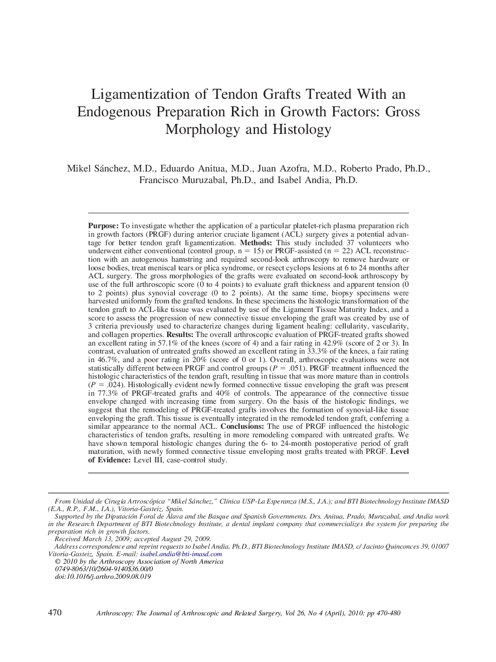
PurposeTo investigate whether the application of a particular platelet-rich plasma preparation rich in growth factors (PRGF) during anterior cruciate ligament (ACL) surgery gives a potential advantage for better tendon graft ligamentization.MethodsThis study included 37 volunteers who underwent either conventional (control group, n = 15) or PRGF-assisted (n = 22) ACL reconstruction with an autogenous hamstring and required second-look arthroscopy to remove hardware or loose bodies, treat meniscal tears or plica syndrome, or resect cyclops lesions at 6 to 24 months after ACL surgery. The gross morphologies of the grafts were evaluated on second-look arthroscopy by use of the full arthroscopic score (0 to 4 points) to evaluate graft thickness and apparent tension (0 to 2 points) plus synovial coverage (0 to 2 points). At the same time, biopsy specimens were harvested uniformly from the grafted tendons. In these specimens the histologic transformation of the tendon graft to ACL-like tissue was evaluated by use of the Ligament Tissue Maturity Index, and a score to assess the progression of new connective tissue enveloping the graft was created by use of 3 criteria previously used to characterize changes during ligament healing: cellularity, vascularity, and collagen properties.ResultsThe overall arthroscopic evaluation of PRGF-treated grafts showed an excellent rating in 57.1% of the knees (score of 4) and a fair rating in 42.9% (score of 2 or 3). In contrast, evaluation of untreated grafts showed an excellent rating in 33.3% of the knees, a fair rating in 46.7%, and a poor rating in 20% (score of 0 or 1). Overall, arthroscopic evaluations were not statistically different between PRGF and control groups (P = .051). PRGF treatment influenced the histologic characteristics of the tendon graft, resulting in tissue that was more mature than in controls (P = .024). Histologically evident newly formed connective tissue enveloping the graft was present in 77.3% of PRGF-treated grafts and 40% of controls. The appearance of the connective tissue envelope changed with increasing time from surgery. On the basis of the histologic findings, we suggest that the remodeling of PRGF-treated grafts involves the formation of synovial-like tissue enveloping the graft. This tissue is eventually integrated in the remodeled tendon graft, conferring a similar appearance to the normal ACL.ConclusionsThe use of PRGF influenced the histologic characteristics of tendon grafts, resulting in more remodeling compared with untreated grafts. We have shown temporal histologic changes during the 6- to 24-month postoperative period of graft maturation, with newly formed connective tissue enveloping most grafts treated with PRGF.Level of EvidenceLevel III, case-control study.
