| Article ID | Journal | Published Year | Pages | File Type |
|---|---|---|---|---|
| 4047505 | Arthroscopy: The Journal of Arthroscopic & Related Surgery | 2007 | 7 Pages |
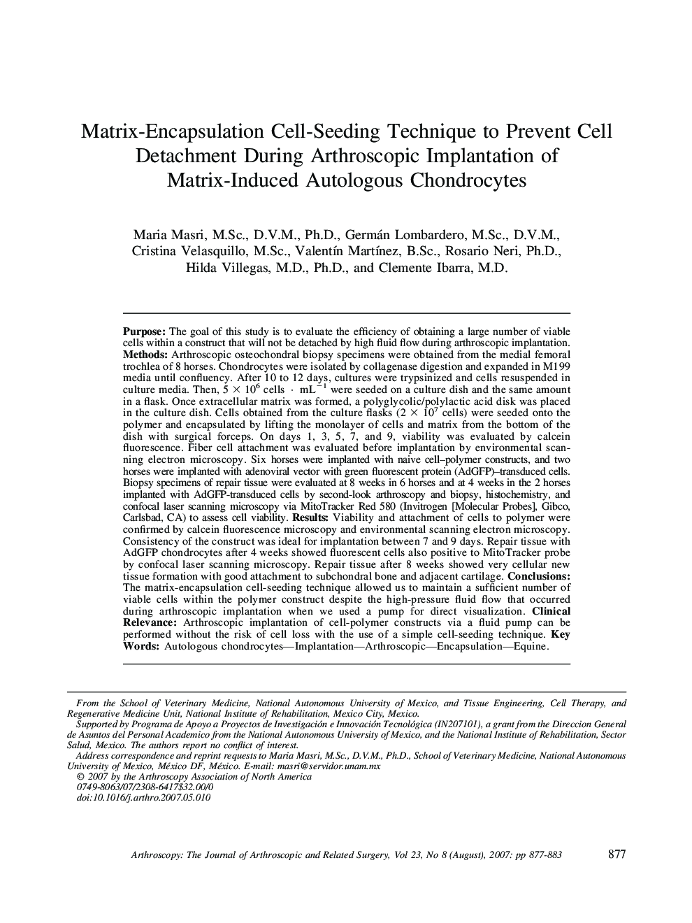
Purpose: The goal of this study is to evaluate the efficiency of obtaining a large number of viable cells within a construct that will not be detached by high fluid flow during arthroscopic implantation. Methods: Arthroscopic osteochondral biopsy specimens were obtained from the medial femoral trochlea of 8 horses. Chondrocytes were isolated by collagenase digestion and expanded in M199 media until confluency. After 10 to 12 days, cultures were trypsinized and cells resuspended in culture media. Then, 5 × 10⁶ cells · mL−1 were seeded on a culture dish and the same amount in a flask. Once extracellular matrix was formed, a polyglycolic/polylactic acid disk was placed in the culture dish. Cells obtained from the culture flasks (2 × 10⁷ cells) were seeded onto the polymer and encapsulated by lifting the monolayer of cells and matrix from the bottom of the dish with surgical forceps. On days 1, 3, 5, 7, and 9, viability was evaluated by calcein fluorescence. Fiber cell attachment was evaluated before implantation by environmental scanning electron microscopy. Six horses were implanted with naive cell–polymer constructs, and two horses were implanted with adenoviral vector with green fluorescent protein (AdGFP)–transduced cells. Biopsy specimens of repair tissue were evaluated at 8 weeks in 6 horses and at 4 weeks in the 2 horses implanted with AdGFP-transduced cells by second-look arthroscopy and biopsy, histochemistry, and confocal laser scanning microscopy via MitoTracker Red 580 (Invitrogen [Molecular Probes], Gibco, Carlsbad, CA) to assess cell viability. Results: Viability and attachment of cells to polymer were confirmed by calcein fluorescence microscopy and environmental scanning electron microscopy. Consistency of the construct was ideal for implantation between 7 and 9 days. Repair tissue with AdGFP chondrocytes after 4 weeks showed fluorescent cells also positive to MitoTracker probe by confocal laser scanning microscopy. Repair tissue after 8 weeks showed very cellular new tissue formation with good attachment to subchondral bone and adjacent cartilage. Conclusions: The matrix-encapsulation cell-seeding technique allowed us to maintain a sufficient number of viable cells within the polymer construct despite the high-pressure fluid flow that occurred during arthroscopic implantation when we used a pump for direct visualization. Clinical Relevance: Arthroscopic implantation of cell-polymer constructs via a fluid pump can be performed without the risk of cell loss with the use of a simple cell-seeding technique.
