| Article ID | Journal | Published Year | Pages | File Type |
|---|---|---|---|---|
| 4168152 | The Journal of Pediatrics | 2010 | 5 Pages |
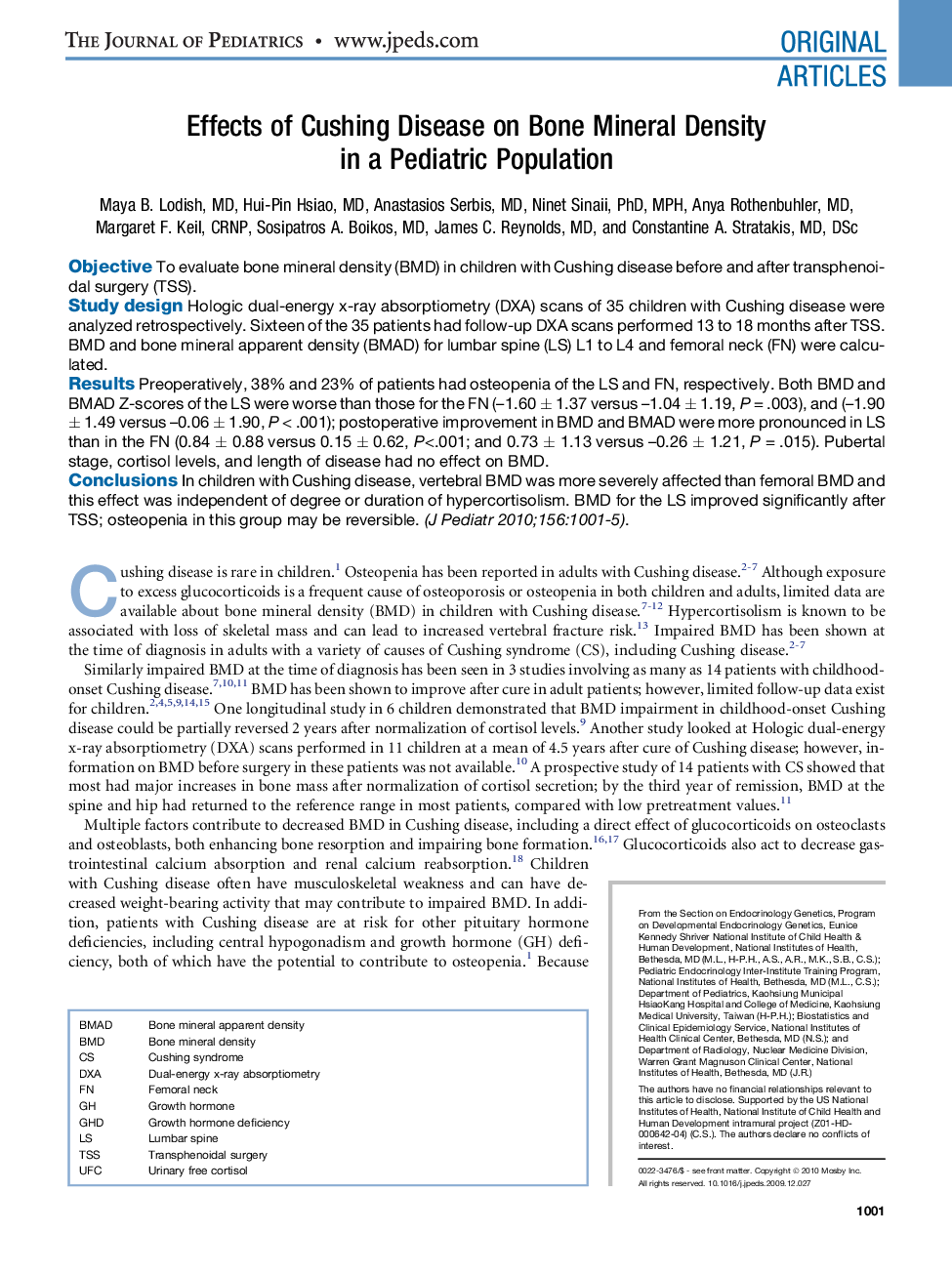
ObjectiveTo evaluate bone mineral density (BMD) in children with Cushing disease before and after transphenoidal surgery (TSS).Study designHologic dual-energy x-ray absorptiometry (DXA) scans of 35 children with Cushing disease were analyzed retrospectively. Sixteen of the 35 patients had follow-up DXA scans performed 13 to 18 months after TSS. BMD and bone mineral apparent density (BMAD) for lumbar spine (LS) L1 to L4 and femoral neck (FN) were calculated.ResultsPreoperatively, 38% and 23% of patients had osteopenia of the LS and FN, respectively. Both BMD and BMAD Z-scores of the LS were worse than those for the FN (–1.60 ± 1.37 versus –1.04 ± 1.19, P = .003), and (–1.90 ± 1.49 versus –0.06 ± 1.90, P < .001); postoperative improvement in BMD and BMAD were more pronounced in LS than in the FN (0.84 ± 0.88 versus 0.15 ± 0.62, P<.001; and 0.73 ± 1.13 versus –0.26 ± 1.21, P = .015). Pubertal stage, cortisol levels, and length of disease had no effect on BMD.ConclusionsIn children with Cushing disease, vertebral BMD was more severely affected than femoral BMD and this effect was independent of degree or duration of hypercortisolism. BMD for the LS improved significantly after TSS; osteopenia in this group may be reversible.
