| Article ID | Journal | Published Year | Pages | File Type |
|---|---|---|---|---|
| 4233233 | Journal of Medical Ultrasound | 2006 | 7 Pages |
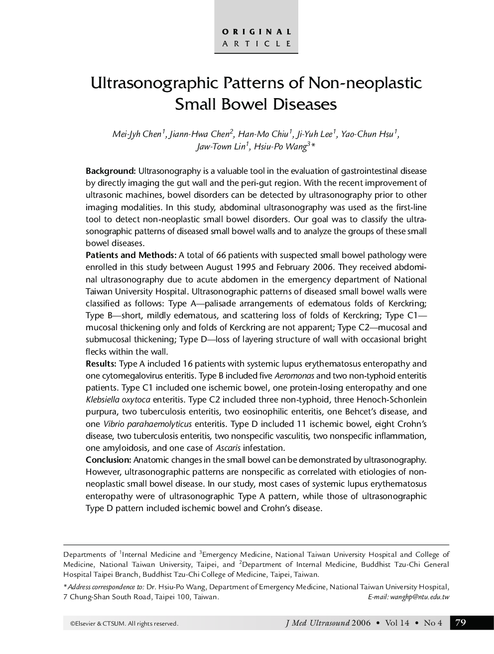
BackgroundUltrasonography is a valuable tool in the evaluation of gastrointestinal disease by directly imaging the gut wall and the perigut region. With the recent improvement of ultrasonic machines, bowel disorders can be detected by ultrasonography prior to other imaging modalities. In this study, abdominal ultrasonography was used as the first-line tool to detect non-neoplastic small bowel disorders. Our goal was to classify the ultrasonographic patterns of diseased small bowel walls and to analyze the groups of these small bowel diseases.Patients and MethodsA total of 66 patients with suspected small bowel pathology were enrolled in this study between August 1995 and February 2006. They received abdominal ultrasonography due to acute abdomen in the emergency department of National Taiwan University Hospital. Ultrasonographic patterns of diseased small bowel walls were classified as follows: Type A—palisade arrangements of edematous folds of Kerckring; Type B—short, mildly edematous, and scattering loss of folds of Kerckring; Type C1—mucosal thickening only and folds of Kerckring are not apparent; Type C2—mucosal and submucosal thickening; Type D—loss of layering structure of wall with occasional bright flecks within the wall.ResultsType A included 16 patients with systemic lupus erythematosus enteropathy and one cytomegalovirus enteritis. Type B included five Aeromonas and two non-typhoid enteritis patients. Type C1 included one ischemic bowel, one protein-losing enteropathy and one Klebsiella oxytoca enteritis. Type C2 included three non-typhoid, three Henoch-Schonlein purpura, two tuberculosis enteritis, two eosinophilic enteritis, one Behcet's disease, and one Vibrio parahaemolyticus enteritis. Type D included 11 ischemic bowel, eight Crohn's disease, two tuberculosis enteritis, two nonspecific vasculitis, two nonspecific inflammation, one amyloidosis, and one case of Ascaris infestation.ConclusionAnatomic changes in the small bowel can be demonstrated by ultrasonography. However, ultrasonographic patterns are nonspecific as correlated with etiologies of non-neoplastic small bowel disease. In our study, most cases of systemic lupus erythematosus enteropathy were of ultrasonographic Type A pattern, while those of ultrasonographic Type D pattern included ischemic bowel and Crohn's disease.
