| Article ID | Journal | Published Year | Pages | File Type |
|---|---|---|---|---|
| 4248192 | Radiology Case Reports | 2011 | 4 Pages |
Abstract
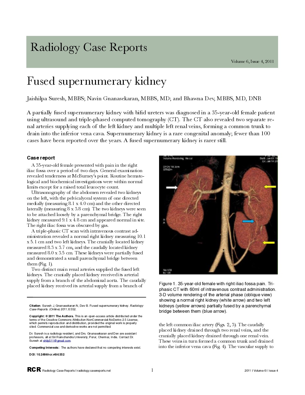
A partially fused supernumerary kidney with bifid ureters was diagnosed in a 35-year-old female patient using ultrasound and triple-phased computed tomography (CT). The CT also revealed two separate renal arteries supplying each of the left kidney and multiple left renal veins, forming a common trunk to drain into the inferior vena cava. Supernumerary kidney is a rare congenital anomaly; fewer than 100 cases have been reported over the years. A fused supernumerary kidney is rarer still.
Related Topics
Health Sciences
Medicine and Dentistry
Radiology and Imaging
Authors
Jaishilpa MBBS, Navin MBBS, MD, Bhawna MBBS, MD, DNB,
