| Article ID | Journal | Published Year | Pages | File Type |
|---|---|---|---|---|
| 4248326 | Radiology Case Reports | 2009 | 6 Pages |
Abstract
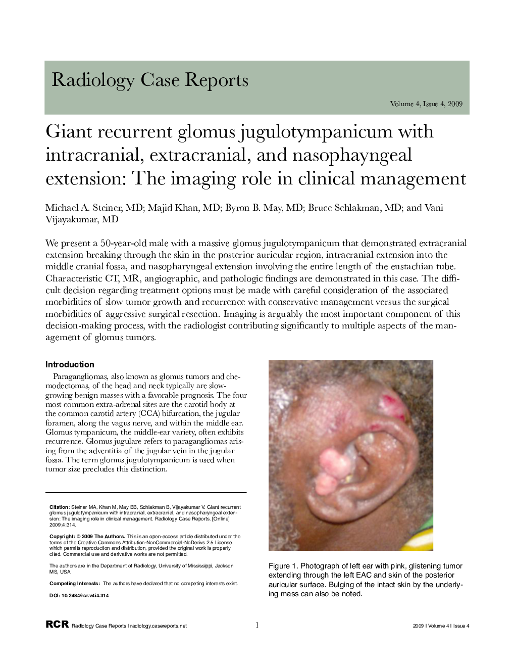
We present a 50-year-old male with a massive glomus jugulotympanicum that demonstrated extracranial extension breaking through the skin in the posterior auricular region, intracranial extension into the middle cranial fossa, and nasopharyngeal extension involving the entire length of the eustachian tube. Characteristic CT, MR, angiographic, and pathologic findings are demonstrated in this case. The difficult decision regarding treatment options must be made with careful consideration of the associated morbidities of slow tumor growth and recurrence with conservative management versus the surgical morbidities of aggressive surgical resection. Imaging is arguably the most important component of this decision-making process, with the radiologist contributing significantly to multiple aspects of the management of glomus tumors.
Keywords
Related Topics
Health Sciences
Medicine and Dentistry
Radiology and Imaging
Authors
Michael A. MD, Majid MD, Byron B. MD, Bruce MD, Vani MD,
