| Article ID | Journal | Published Year | Pages | File Type |
|---|---|---|---|---|
| 4250728 | Revista Española de Medicina Nuclear e Imagen Molecular (English Edition) | 2012 | 4 Pages |
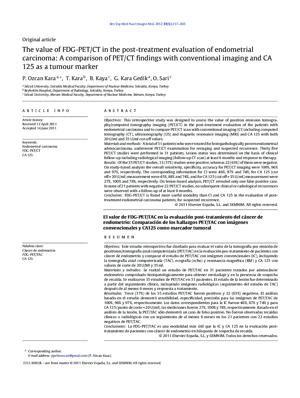
ObjectivesThis retrospective study was designed to assess the value of positron emission tomography/computed tomography imaging (PET/CT) in the post-treatment evaluation of the patients with endometrial carcinoma and to compare PET/CT scan with conventional imaging (CI) including computed tomography (CT), ultrasonography (US) and magnetic resonance imaging (MRI) and CA 125 with both 20 U/ml and 35 U/ml cut-off values.Materials and methodsA total of 31 patients who were treated for histopathologically proven endometrial adenocarcinoma, underwent PET/CT examination for restaging and suspected recurrence. Thirty five PET/CT studies were performed in 31 patients. Lesion status was determined on the basis of clinical follow-up including radiological imaging (follow-up CT scan) at least 6 months and response to therapy.ResultsOf the 35 PET/CT studies, 13 (37%) studies were positive, whereas 22 (63%) of them were negative. On study-based analysis the overall sensitivity, specificity, accuracy for PET/CT imaging were 100%, 96% and 97%, respectively. The corresponding information for CI were 46%, 87% and 74%, for CA 125 (cut off = 20 U/ml) measurement were 45%, 88% and 74%, and for CA 125 (cut off = 35 U/ml) measurement were 27%, 100% and 78%, respectively. On lesion-based analysis, PET/CT revealed only one false positive case. In none of 21 patients with negative 22 PET/CT studies, no subsequent clinical or radiological recurrences were observed with a follow-up of at least 6 months.ConclusionFDG-PET/CT is found more useful modality than CI and CA 125 in the evaluation of post-treatment endometrial carcinoma patients, for suspected recurrence.
ResumenObjetivosEste estudio retrospectivo fue diseñado para evaluar el valor de la tomografía por emisión de positrones/tomografía axial computerizada (PET/TAC) en la evaluación pos-tratamiento de pacientes con cáncer de endometrio y comparar el estudio de PET/TAC con imágenes convencionales (IC), incluyendo la tomografía axial computerizada (TAC), ecografía (echo) y resonancia magnética (RM) y CA 125 con valores de corte de 20 U/Ml y 35 ml.Materiales y métodosSe realizó un estudio de PET/TAC en 31 pacientes tratados por adenocáncer endometrio comprobado histopatologicamente para obtener reestadiaje y en la presencia de sospecha de recaída. Se realizaron 35 estudios de PET/TAC en 31 pacientes. El estado de la lesión fue determinado a partir del seguimiento clínico, incluyendo imágenes radiológicas (seguimiento del estudio de TAC) después de al menos 6 meses y respuesta a tratamiento.ResultadosTrece (37%) de los 35 estudios PET/TAC fueron positivos y 22 (63%) negativos. El análisis basado en el estudio demostró sensibilidad, especificidad, precisión para las imágenes de PET/TAC de 100%, 96% y 97%, respectivamente. Los datos correspondientes para la IC fueron 46%, 87% y 74% y para CA 125 (punto de corte = 20 U/ml), las mediciones fueron 27%, 100% y 78%, respectivamente. Basado en el análisis de la lesión, la PET/TAC sólo demostró un caso de falso positivo. No fueron observadas recaídas clínicas o radiológicas con un seguimiento de al menos 6 meses en los 21 pacientes con 22 estudios negativos de PET/TAC.ConclusionesLa FDG-PET/TAC es una modalidad más útil que la IC y CA 125 en la evaluación post-tratamiento de pacientes con cáncer de endometrio en búsqueda de sospecha de recaída.
