| Article ID | Journal | Published Year | Pages | File Type |
|---|---|---|---|---|
| 5663577 | Acta Haematologica Polonica | 2017 | 6 Pages |
Abstract
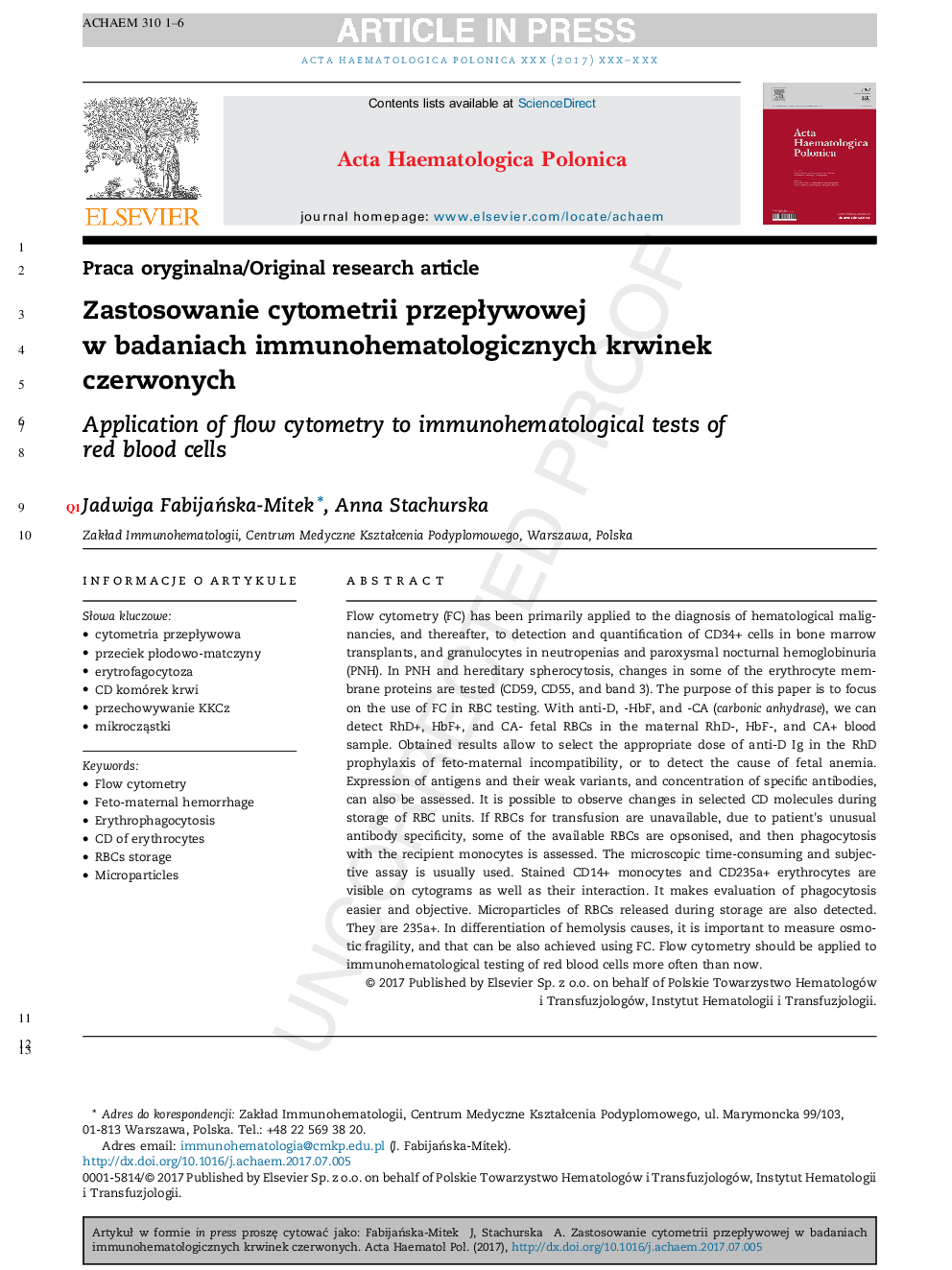
Flow cytometry (FC) has been primarily applied to the diagnosis of hematological malignancies, and thereafter, to detection and quantification of CD34+ cells in bone marrow transplants, and granulocytes in neutropenias and paroxysmal nocturnal hemoglobinuria (PNH). In PNH and hereditary spherocytosis, changes in some of the erythrocyte membrane proteins are tested (CD59, CD55, and band 3). The purpose of this paper is to focus on the use of FC in RBC testing. With anti-D, -HbF, and -CA (carbonic anhydrase), we can detect RhD+, HbF+, and CA- fetal RBCs in the maternal RhD-, HbF-, and CA+ blood sample. Obtained results allow to select the appropriate dose of anti-D Ig in the RhD prophylaxis of feto-maternal incompatibility, or to detect the cause of fetal anemia. Expression of antigens and their weak variants, and concentration of specific antibodies, can also be assessed. It is possible to observe changes in selected CD molecules during storage of RBC units. If RBCs for transfusion are unavailable, due to patient's unusual antibody specificity, some of the available RBCs are opsonised, and then phagocytosis with the recipient monocytes is assessed. The microscopic time-consuming and subjective assay is usually used. Stained CD14+ monocytes and CD235a+ erythrocytes are visible on cytograms as well as their interaction. It makes evaluation of phagocytosis easier and objective. Microparticles of RBCs released during storage are also detected. They are 235a+. In differentiation of hemolysis causes, it is important to measure osmotic fragility, and that can be also achieved using FC. Flow cytometry should be applied to immunohematological testing of red blood cells more often than now.
Related Topics
Life Sciences
Immunology and Microbiology
Immunology
Authors
Jadwiga FabijaÅska-Mitek, Anna Stachurska,
