| Article ID | Journal | Published Year | Pages | File Type |
|---|---|---|---|---|
| 5936387 | The American Journal of Pathology | 2009 | 10 Pages |
Abstract
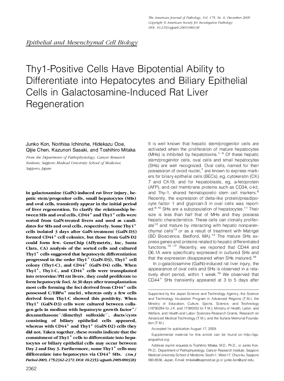
In galactosamine (GalN)-induced rat liver injury, hepatic stem/progenitor cells, small hepatocytes (SHs) and oval cells, transiently appear in the initial period of liver regeneration. To clarify the relationship between SHs and oval cells, CD44+ and Thy1+ cells were sorted from GalN-treated livers and used as candidates for SHs and oval cells, respectively. Some Thy1+ cells isolated 3 days after GalN-treatment (GalN-D3) formed CD44+ cell colonies, but those from GalN-D2 could form few. GeneChip (Affymetrix, Inc, Santa Clara, CA) analysis of the sorted cells and cultured Thy1+ cells suggested that hepatocytic differentiation progressed in the order Thy1+ (GalN-D3), Thy1+ cell colony (Thy1-C), and CD44+ (GalN-D4) cells. When Thy1+, Thy1-C, and CD44+ cells were transplanted into retrorsine/PH rat livers, they could proliferate to form hepatocytic foci. At 30 days after transplantation most cells forming the foci derived from CD44+ cells possessed C/EBPα+ nuclei, whereas only a few cells derived from Thy1-C showed this positivity. When Thy1+ (GalN-D3) cells were cultured between collagen gels in medium with hepatocyte growth factor+/dexamethasoneâ/dimethyl sulfoxideâ, ducts/cysts consisting of biliary epithelial cells appeared, whereas with CD44+ and Thy1+ (GalN-D2) cells they did not. Taken together, these results indicate that the commitment of Thy1+ cells to differentiate into hepatocytes or biliary epithelial cells may occur between Day 2 and Day 3. Furthermore, some Thy1+ cells may differentiate into hepatocytes via CD44+ SHs.
Related Topics
Health Sciences
Medicine and Dentistry
Cardiology and Cardiovascular Medicine
Authors
Junko Kon, Norihisa Ichinohe, Hidekazu Ooe, Qijie Chen, Kazunori Sasaki, Toshihiro Mitaka,
