| Article ID | Journal | Published Year | Pages | File Type |
|---|---|---|---|---|
| 6056306 | Oral Surgery, Oral Medicine, Oral Pathology and Oral Radiology | 2014 | 8 Pages |
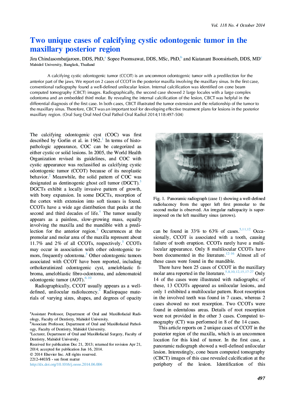
A calcifying cystic odontogenic tumor (CCOT) is an uncommon odontogenic tumor with a predilection for the anterior part of the jaws. We report on 2 cases of CCOT in the posterior maxilla involving the maxillary sinus. In the first case, conventional radiography found a well-defined unilocular lesion. Internal calcification was identified on cone beam computed tomography (CBCT) images. Radiographically, the second case showed 2 large locules with a large complex odontoma and an embedded third molar. By revealing the internal calcification of the lesion, CBCT was helpful in the differential diagnosis of the first case. In both cases, CBCT illustrated the tumor extension and the relationship of the tumor to the maxillary sinus. Therefore, CBCT was an important tool for developing effective treatment plans for lesions in the posterior maxillary region.
