| Article ID | Journal | Published Year | Pages | File Type |
|---|---|---|---|---|
| 6144932 | American Journal of Obstetrics and Gynecology | 2014 | 11 Pages |
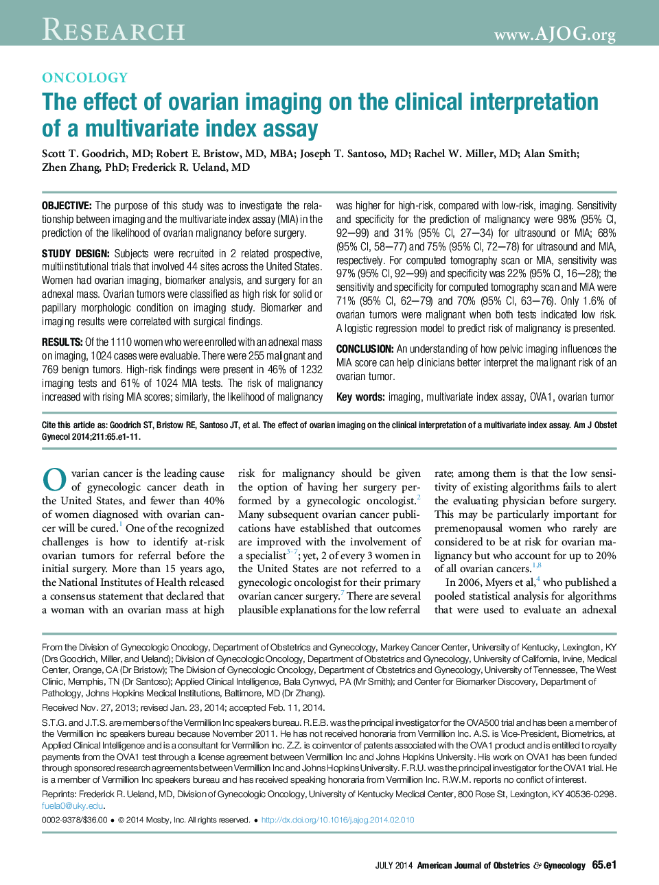
ObjectiveThe purpose of this study was to investigate the relationship between imaging and the multivariate index assay (MIA) in the prediction of the likelihood of ovarian malignancy before surgery.Study DesignSubjects were recruited in 2 related prospective, multiinstitutional trials that involved 44 sites across the United States. Women had ovarian imaging, biomarker analysis, and surgery for an adnexal mass. Ovarian tumors were classified as high risk for solid or papillary morphologic condition on imaging study. Biomarker and imaging results were correlated with surgical findings.ResultsOf the 1110 women who were enrolled with an adnexal mass on imaging, 1024 cases were evaluable. There were 255 malignant and 769 benign tumors. High-risk findings were present in 46% of 1232 imaging tests and 61% of 1024 MIA tests. The risk of malignancy increased with rising MIA scores; similarly, the likelihood of malignancy was higher for high-risk, compared with low-risk, imaging. Sensitivity and specificity for the prediction of malignancy were 98% (95% CI, 92-99) and 31% (95% CI, 27-34) for ultrasound or MIA; 68% (95%Â CI, 58-77) and 75% (95% CI, 72-78) for ultrasound and MIA, respectively. For computed tomography scan or MIA, sensitivity was 97% (95% CI, 92-99) and specificity was 22% (95% CI, 16-28); the sensitivity and specificity for computed tomography scan and MIA were 71% (95% CI, 62-79) and 70% (95% CI, 63-76). Only 1.6% of ovarian tumors were malignant when both tests indicated low risk. AÂ logistic regression model to predict risk of malignancy is presented.ConclusionAn understanding of how pelvic imaging influences the MIA score can help clinicians better interpret the malignant risk of an ovarian tumor.
