| Article ID | Journal | Published Year | Pages | File Type |
|---|---|---|---|---|
| 6179280 | Fertility and Sterility | 2013 | 9 Pages |
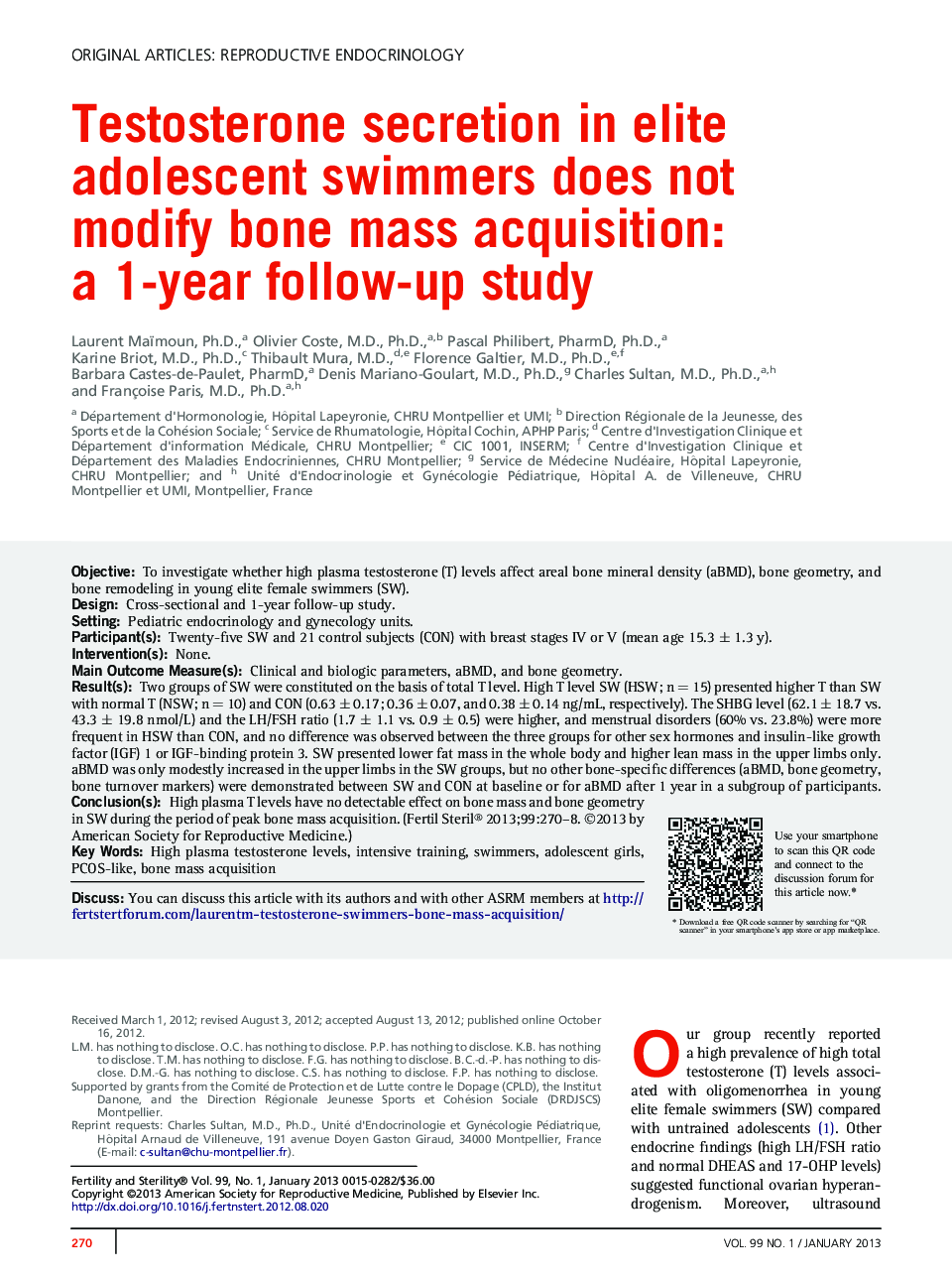
ObjectiveTo investigate whether high plasma testosterone (T) levels affect areal bone mineral density (aBMD), bone geometry, and bone remodeling in young elite female swimmers (SW).DesignCross-sectional and 1-year follow-up study.SettingPediatric endocrinology and gynecology units.Participant(s)Twenty-five SW and 21 control subjects (CON) with breast stages IV or V (mean age 15.3 ± 1.3 y).Intervention(s)None.Main Outcome Measure(s)Clinical and biologic parameters, aBMD, and bone geometry.Result(s)Two groups of SW were constituted on the basis of total T level. High T level SW (HSW; n = 15) presented higher T than SW with normal T (NSW; n = 10) and CON (0.63 ± 0.17; 0.36 ± 0.07, and 0.38 ± 0.14 ng/mL, respectively). The SHBG level (62.1 ± 18.7 vs. 43.3 ± 19.8 nmol/L) and the LH/FSH ratio (1.7 ± 1.1 vs. 0.9 ± 0.5) were higher, and menstrual disorders (60% vs. 23.8%) were more frequent in HSW than CON, and no difference was observed between the three groups for other sex hormones and insulin-like growth factor (IGF) 1 or IGF-binding protein 3. SW presented lower fat mass in the whole body and higher lean mass in the upper limbs only. aBMD was only modestly increased in the upper limbs in the SW groups, but no other bone-specific differences (aBMD, bone geometry, bone turnover markers) were demonstrated between SW and CON at baseline or for aBMD after 1 year in a subgroup of participants.Conclusion(s)High plasma T levels have no detectable effect on bone mass and bone geometry in SW during the period of peak bone mass acquisition.
