| Article ID | Journal | Published Year | Pages | File Type |
|---|---|---|---|---|
| 6181033 | Fertility and Sterility | 2015 | 11 Pages |
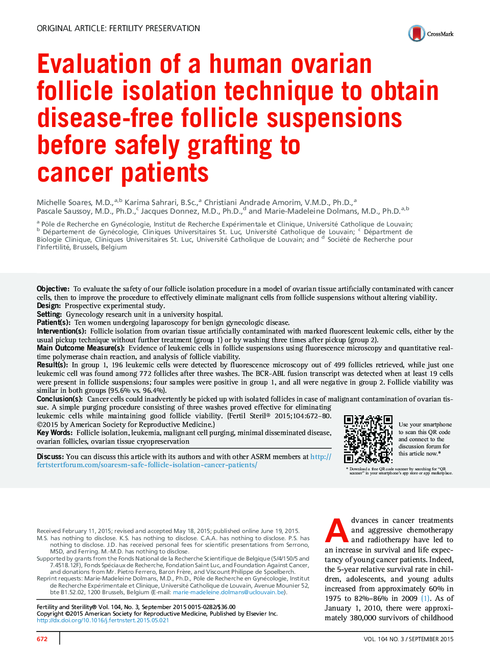
ObjectiveTo evaluate the safety of our follicle isolation procedure in a model of ovarian tissue artificially contaminated with cancer cells, then to improve the procedure to effectively eliminate malignant cells from follicle suspensions without altering viability.DesignProspective experimental study.SettingGynecology research unit in a university hospital.Patient(s)Ten women undergoing laparoscopy for benign gynecologic disease.Intervention(s)Follicle isolation from ovarian tissue artificially contaminated with marked fluorescent leukemic cells, either by the usual pickup technique without further treatment (group 1) or by washing three times after pickup (group 2).Main Outcome Measure(s)Evidence of leukemic cells in follicle suspensions using fluorescence microscopy and quantitative real-time polymerase chain reaction, and analysis of follicle viability.Result(s)In group 1, 196 leukemic cells were detected by fluorescence microscopy out of 499 follicles retrieved, while just one leukemic cell was found among 772 follicles after three washes. The BCR-ABL fusion transcript was detected when at least 19 cells were present in follicle suspensions; four samples were positive in group 1, and all were negative in group 2. Follicle viability was similar in both groups (95.6% vs. 96.4%).Conclusion(s)Cancer cells could inadvertently be picked up with isolated follicles in case of malignant contamination of ovarian tissue. A simple purging procedure consisting of three washes proved effective for eliminating leukemic cells while maintaining good follicle viability.
