| Article ID | Journal | Published Year | Pages | File Type |
|---|---|---|---|---|
| 6271785 | Neuroscience | 2015 | 11 Pages |
Abstract
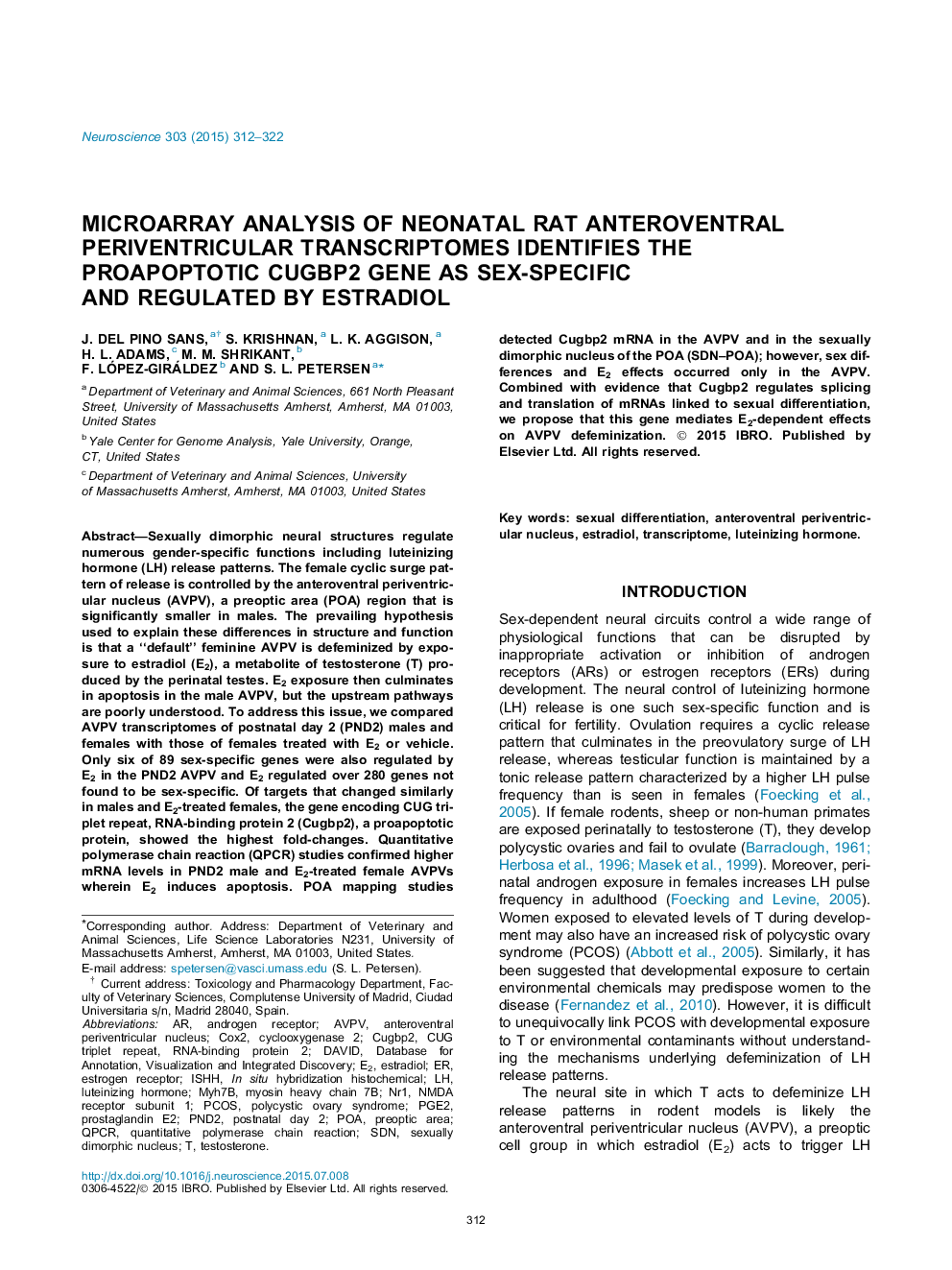
Sexually dimorphic neural structures regulate numerous gender-specific functions including luteinizing hormone (LH) release patterns. The female cyclic surge pattern of release is controlled by the anteroventral periventricular nucleus (AVPV), a preoptic area (POA) region that is significantly smaller in males. The prevailing hypothesis used to explain these differences in structure and function is that a “default” feminine AVPV is defeminized by exposure to estradiol (E2), a metabolite of testosterone (T) produced by the perinatal testes. E2 exposure then culminates in apoptosis in the male AVPV, but the upstream pathways are poorly understood. To address this issue, we compared AVPV transcriptomes of postnatal day 2 (PND2) males and females with those of females treated with E2 or vehicle. Only six of 89 sex-specific genes were also regulated by E2 in the PND2 AVPV and E2 regulated over 280 genes not found to be sex-specific. Of targets that changed similarly in males and E2-treated females, the gene encoding CUG triplet repeat, RNA-binding protein 2 (Cugbp2), a proapoptotic protein, showed the highest fold-changes. Quantitative polymerase chain reaction (QPCR) studies confirmed higher mRNA levels in PND2 male and E2-treated female AVPVs wherein E2 induces apoptosis. POA mapping studies detected Cugbp2 mRNA in the AVPV and in the sexually dimorphic nucleus of the POA (SDN-POA); however, sex differences and E2 effects occurred only in the AVPV. Combined with evidence that Cugbp2 regulates splicing and translation of mRNAs linked to sexual differentiation, we propose that this gene mediates E2-dependent effects on AVPV defeminization.
Keywords
SDNqPCRPOAPGE2PCOScox2NMDA receptor subunit 1AVPVISHHNR1EstradiolTranscriptometestosteroneSexual differentiationDAVIDpostnatal day 2Polycystic ovary syndromecyclooxygenase 2Preoptic areaSexually dimorphic nucleusanteroventral periventricular nucleusluteinizing hormonequantitative polymerase chain reactiondatabase for annotation, visualization and integrated discoveryProstaglandin E2Androgen ReceptorEstrogen receptor
Related Topics
Life Sciences
Neuroscience
Neuroscience (General)
Authors
J. Del Pino Sans, S. Krishnan, L.K. Aggison, H.L. Adams, M.M. Shrikant, F. López-Giráldez, S.L. Petersen,
