| Article ID | Journal | Published Year | Pages | File Type |
|---|---|---|---|---|
| 6972107 | Journal of Hazardous Materials | 2013 | 6 Pages |
Abstract
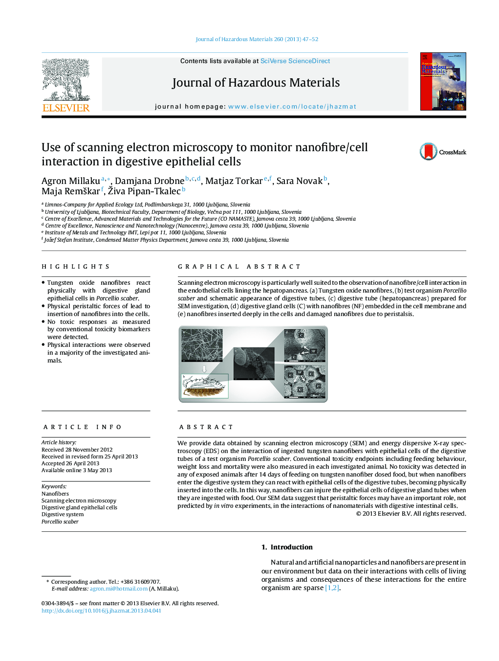
Scanning electron microscopy is particularly well suited to the observation of nanofibre/cell interaction in the endothelial cells lining the hepatopancreas. (a) Tungsten oxide nanofibres, (b) test organism Porcellio scaber and schematic appearance of digestive tubes, (c) digestive tube (hepatopancreas) prepared for SEM investigation, (d) digestive gland cells (C) with nanofibres (NF) embedded in the cell membrane and (e) nanofibres inserted deeply in the cells and damaged nanofibres due to peristalsis.
Related Topics
Physical Sciences and Engineering
Chemical Engineering
Chemical Health and Safety
Authors
Agron Millaku, Damjana Drobne, Matjaz Torkar, Sara Novak, Maja Remškar, Živa Pipan-Tkalec,
