| Article ID | Journal | Published Year | Pages | File Type |
|---|---|---|---|---|
| 7290893 | Human Movement Science | 2018 | 8 Pages |
Abstract
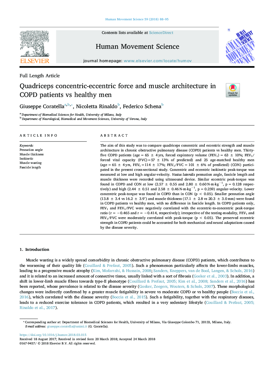
The aim of this study was to compare quadriceps concentric and eccentric strength and muscle architecture in chronic obstructive pulmonary disease (COPD) patients vs healthy men. Thirty-five COPD patients (ageâ¯=â¯65â¯Â±â¯4â¯yrs, forced expiratory volume (FEV1)â¯=â¯63â¯Â±â¯10%; FEV1/forced vital capacity (FVC)=57â¯Â±â¯13% of predicted) and 25 age-matched healthy men (ageâ¯=â¯65â¯Â±â¯4â¯yrs, FEV1=114â¯Â±â¯17%; FEV1/FVCâ¯=â¯101â¯Â±â¯6% of predicted) (CON) participated in the present cross-sectional study. Concentric and eccentric isokinetic peak-torque was measured at low and high angular-velocity. Vastus lateralis pennation angle, fascicle length and muscle thickness were recorded using ultrasound device. Similar eccentric peak-torque was found in COPD and CON at low (2.57â¯Â±â¯0.55 and 2.80â¯Â±â¯0.60â¯Nâ
mâ
kgâ1, pâ¯=â¯0.128 respectively) and high (2.44â¯Â±â¯0.51 and 2.58â¯Â±â¯0.46â¯Nâ
mâ
kgâ1, pâ¯=â¯0.259) angular-velocity. Lower concentric peak-torque was found in COPD than in CON (pâ¯<â¯0.05). Smaller pennation angle (13.8â¯Â±â¯3.4 vs 16.2â¯Â±â¯3.9°) and muscle thickness (17.1â¯Â±â¯2.8 vs 20.3â¯Â±â¯3.0â¯mm) were found in COPD patients vs healthy men, with no difference in fascicle length. In COPD patients only, FEV1 and FEV1/FVC were negatively correlated with the eccentric-to-concentric peak-torque ratio (râ¯=â¯â0.465 and râ¯=â¯â0.414, respectively); irrespective of the testing-modality, FEV1 and FEV1/FVC were moderately correlated with peak-torque (pâ¯<â¯0.05). The preserved eccentric strength in COPD patients could be accounted for both mechanical and neural adaptations caused by the disease severity.
Related Topics
Life Sciences
Neuroscience
Cognitive Neuroscience
Authors
Giuseppe Coratella, Nicoletta Rinaldo, Federico Schena,
