| Article ID | Journal | Published Year | Pages | File Type |
|---|---|---|---|---|
| 7319650 | Neuropsychologia | 2015 | 12 Pages |
Abstract
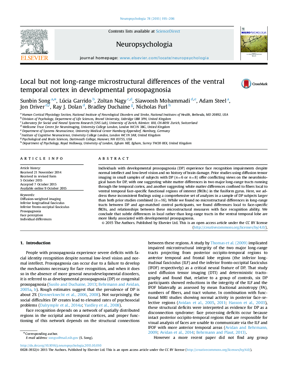
Individuals with developmental prosopagnosia (DP) experience face recognition impairments despite normal intellect and low-level vision and no history of brain damage. Prior studies using diffusion tensor imaging in small samples of subjects with DP (n=6 or n=8) offer conflicting views on the neurobiological bases for DP, with one suggesting white matter differences in two major long-range tracts running through the temporal cortex, and another suggesting white matter differences confined to fibers local to ventral temporal face-specific functional regions of interest (fROIs) in the fusiform gyrus. Here, we address these inconsistent findings using a comprehensive set of analyzes in a sample of DP subjects larger than both prior studies combined (n=16). While we found no microstructural differences in long-range tracts between DP and age-matched control participants, we found differences local to face-specific fROIs, and relationships between these microstructural measures with face recognition ability. We conclude that subtle differences in local rather than long-range tracts in the ventral temporal lobe are more likely associated with developmental prosopagnosia.
Keywords
Related Topics
Life Sciences
Neuroscience
Behavioral Neuroscience
Authors
Sunbin Song, Lúcia Garrido, Zoltan Nagy, Siawoosh Mohammadi, Adam Steel, Jon Driver, Ray J. Dolan, Bradley Duchaine, Nicholas Furl,
