| Article ID | Journal | Published Year | Pages | File Type |
|---|---|---|---|---|
| 7646572 | Revue Francophone des Laboratoires | 2016 | 6 Pages |
Abstract
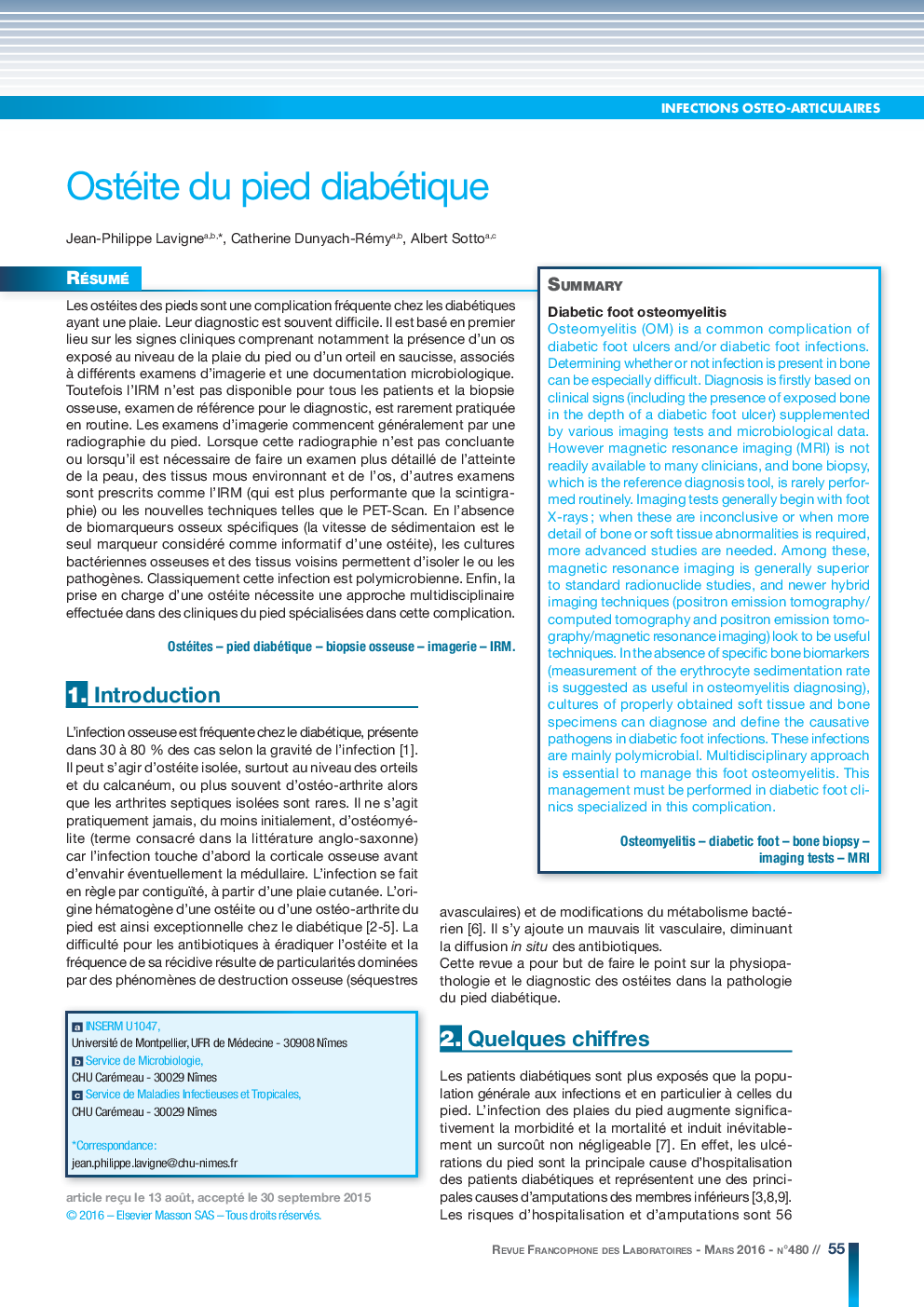
Osteomyelitis (OM) is a common complication of diabetic foot ulcers and/or diabetic foot infections. Determining whether or not infection is present in bone can be especially difficult. Diagnosis is firstly based on clinical signs (including the presence of exposed bone in the depth of a diabetic foot ulcer) supplemented by various imaging tests and microbiological data. However magnetic resonance imaging (MRI) is not readily available to many clinicians, and bone biopsy, which is the reference diagnosis tool, is rarely performed routinely. Imaging tests generally begin with foot X-rays ; when these are inconclusive or when more detail of bone or soft tissue abnormalities is required, more advanced studies are needed. Among these, magnetic resonance imaging is generally superior to standard radionuclide studies, and newer hybrid imaging techniques (positron emission tomography/computed tomography and positron emission tomography/magnetic resonance imaging) look to be useful techniques. In the absence of specific bone biomarkers (measurement of the erythrocyte sedimentation rate is suggested as useful in osteomyelitis diagnosing), cultures of properly obtained soft tissue and bone specimens can diagnose and define the causative pathogens in diabetic foot infections. These infections are mainly polymicrobial. Multidisciplinary approach is essential to manage this foot osteomyelitis. This management must be performed in diabetic foot clinics specialized in this complication.
Related Topics
Physical Sciences and Engineering
Chemistry
Analytical Chemistry
Authors
Jean-Philippe Lavigne, Catherine Dunyach-Rémy, Albert Sotto,
