| Article ID | Journal | Published Year | Pages | File Type |
|---|---|---|---|---|
| 8236874 | International Journal of Radiation Oncology*Biology*Physics | 2009 | 7 Pages |
Abstract
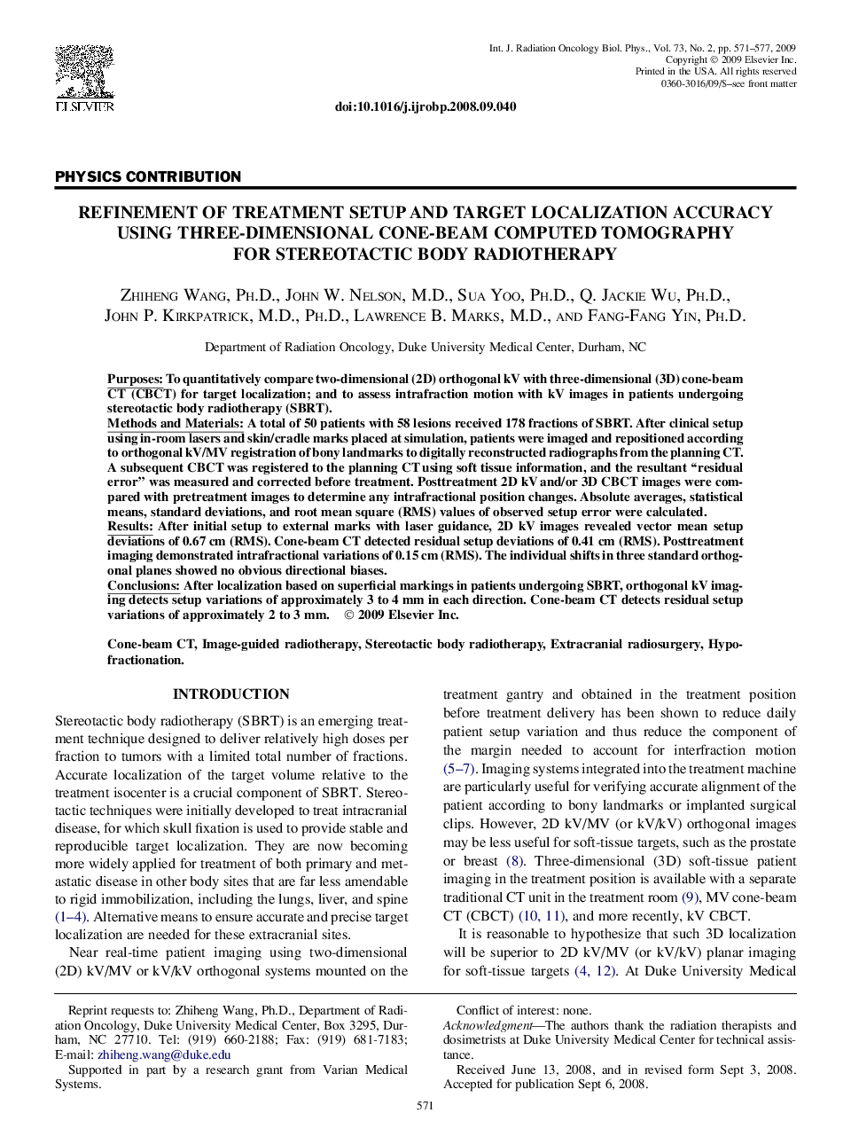
After localization based on superficial markings in patients undergoing SBRT, orthogonal kV imaging detects setup variations of approximately 3 to 4 mm in each direction. Cone-beam CT detects residual setup variations of approximately 2 to 3 mm.
Keywords
Related Topics
Physical Sciences and Engineering
Physics and Astronomy
Radiation
Authors
Zhiheng Ph.D., John W. M.D., Sua Ph.D., Q. Jackie Ph.D., John P. M.D., Ph.D., Lawrence B. M.D., Fang-Fang Ph.D.,
