| Article ID | Journal | Published Year | Pages | File Type |
|---|---|---|---|---|
| 8679094 | Seminars in Thoracic and Cardiovascular Surgery | 2018 | 6 Pages |
Abstract
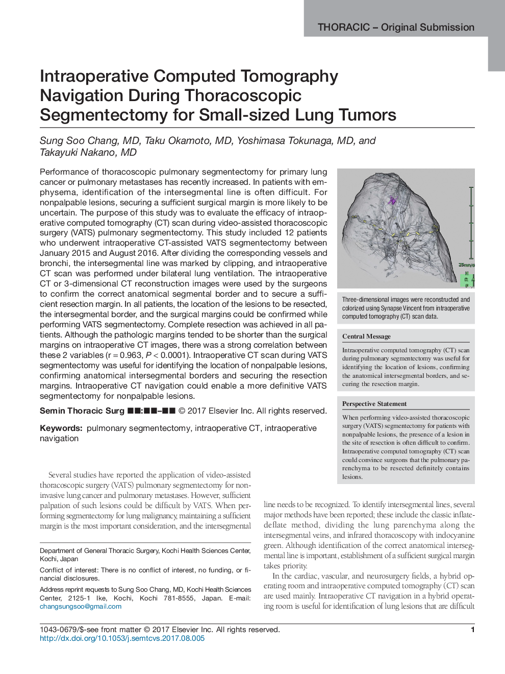
Performance of thoracoscopic pulmonary segmentectomy for primary lung cancer or pulmonary metastases has recently increased. In patients with emphysema, identification of the intersegmental line is often difficult. For nonpalpable lesions, securing a sufficient surgical margin is more likely to be uncertain. The purpose of this study was to evaluate the efficacy of intraoperative computed tomography (CT) scan during video-assisted thoracoscopic surgery (VATS) pulmonary segmentectomy. This study included 12 patients who underwent intraoperative CT-assisted VATS segmentectomy between January 2015 and August 2016. After dividing the corresponding vessels and bronchi, the intersegmental line was marked by clipping, and intraoperative CT scan was performed under bilateral lung ventilation. The intraoperative CT or 3-dimensional CT reconstruction images were used by the surgeons to confirm the correct anatomical segmental border and to secure a sufficient resection margin. In all patients, the location of the lesions to be resected, the intersegmental border, and the surgical margins could be confirmed while performing VATS segmentectomy. Complete resection was achieved in all patients. Although the pathologic margins tended to be shorter than the surgical margins on intraoperative CT images, there was a strong correlation between these 2 variables (râ=â0.963, Pâ<â0.0001). Intraoperative CT scan during VATS segmentectomy was useful for identifying the location of nonpalpable lesions, confirming anatomical intersegmental borders and securing the resection margins. Intraoperative CT navigation could enable a more definitive VATS segmentectomy for nonpalpable lesions.
Related Topics
Health Sciences
Medicine and Dentistry
Cardiology and Cardiovascular Medicine
Authors
Sung Soo MD, Taku MD, Yoshimasa MD, Takayuki MD,
