| Article ID | Journal | Published Year | Pages | File Type |
|---|---|---|---|---|
| 8807557 | Human Pathology | 2018 | 10 Pages |
Abstract
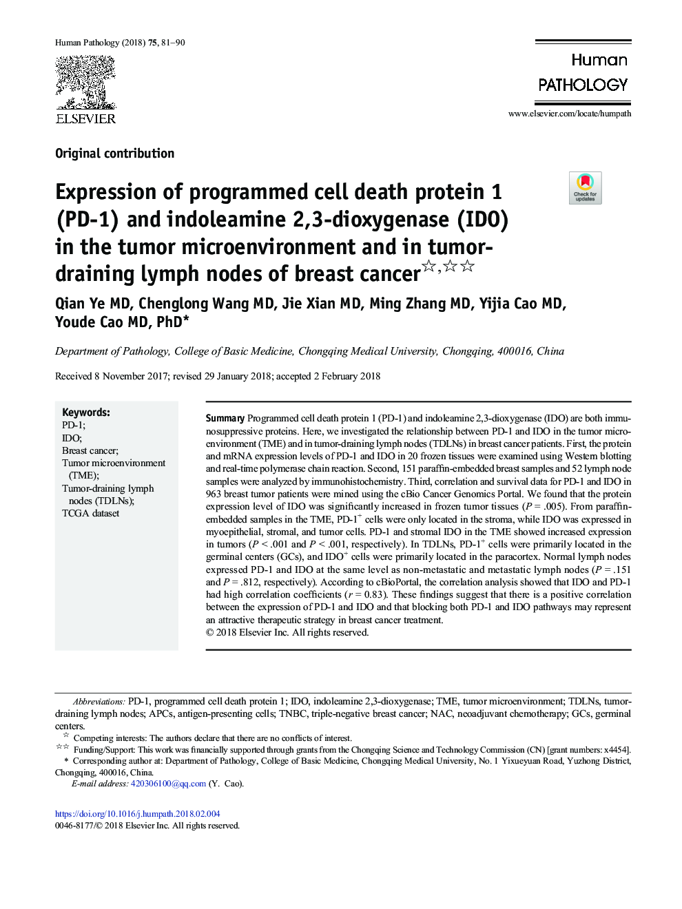
Programmed cell death protein 1 (PD-1) and indoleamine 2,3-dioxygenase (IDO) are both immunosuppressive proteins. Here, we investigated the relationship between PD-1 and IDO in the tumor microenvironment (TME) and in tumor-draining lymph nodes (TDLNs) in breast cancer patients. First, the protein and mRNA expression levels of PD-1 and IDO in 20 frozen tissues were examined using Western blotting and real-time polymerase chain reaction. Second, 151 paraffin-embedded breast samples and 52 lymph node samples were analyzed by immunohistochemistry. Third, correlation and survival data for PD-1 and IDO in 963 breast tumor patients were mined using the cBio Cancer Genomics Portal. We found that the protein expression level of IDO was significantly increased in frozen tumor tissues (P = .005). From paraffin-embedded samples in the TME, PD-1+ cells were only located in the stroma, while IDO was expressed in myoepithelial, stromal, and tumor cells. PD-1 and stromal IDO in the TME showed increased expression in tumors (P< .001 and P < .001, respectively). In TDLNs, PD-1+ cells were primarily located in the germinal centers (GCs), and IDO+ cells were primarily located in the paracortex. Normal lymph nodes expressed PD-1 and IDO at the same level as non-metastatic and metastatic lymph nodes (P = .151 and P = .812, respectively). According to cBioPortal, the correlation analysis showed that IDO and PD-1 had high correlation coefficients (r = 0.83). These findings suggest that there is a positive correlation between the expression of PD-1 and IDO and that blocking both PD-1 and IDO pathways may represent an attractive therapeutic strategy in breast cancer treatment.
Keywords
Related Topics
Health Sciences
Medicine and Dentistry
Pathology and Medical Technology
Authors
Qian MD, Chenglong MD, Jie MD, Ming MD, Yijia MD, Youde MD, PhD,
