| Article ID | Journal | Published Year | Pages | File Type |
|---|---|---|---|---|
| 9089465 | Anaesthesia & Intensive Care Medicine | 2005 | 5 Pages |
Abstract
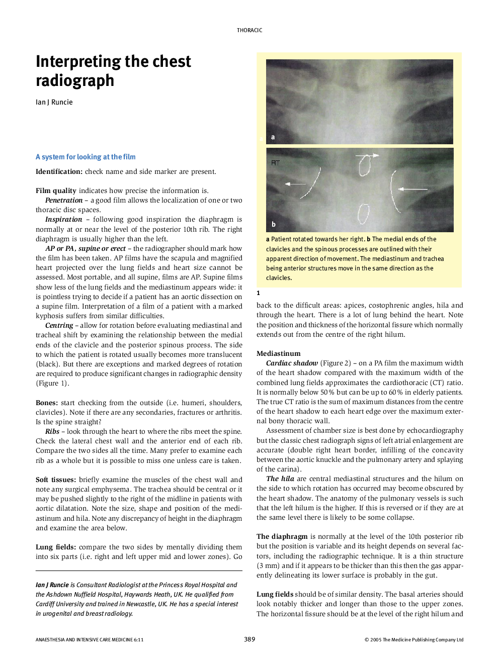
This article provides information about interpreting chest radiographs, with particular attention to the needs of anaesthetists. A routine for looking at each film is suggested, with comments on the assessment of the quality of the film, including rotation and penetration. Mention is made of each anatomical area (bones, soft tissues, mediastinum, lung fields) and the pathological changes seen in each is discussed. The differences between the findings in alveolar and interstitial pathology are emphasized. The lateral film is discussed and special attention is paid to the particular difficulties presented by chest radiographs taken in the ITU. There are discussions on the appearances related to vascular lines and the use of CT in the ITU.
Related Topics
Health Sciences
Medicine and Dentistry
Anesthesiology and Pain Medicine
Authors
Ian J Runcie,
