| Article ID | Journal | Published Year | Pages | File Type |
|---|---|---|---|---|
| 9231182 | Journal of Investigative Dermatology | 2005 | 9 Pages |
Abstract
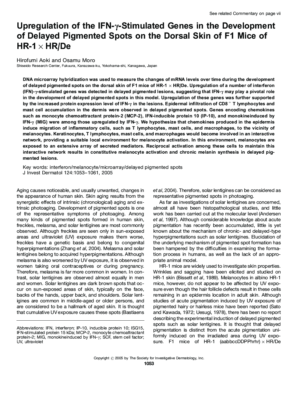
DNA microarray hybridization was used to measure the changes of mRNA levels over time during the development of delayed pigmented spots on the dorsal skin of F1 mice of HR-1 à HR/De. Upregulation of a number of interferon (IFN)-γ-stimulated genes was detected in delayed pigmented lesions, suggesting that IFN-γ may play a pivotal role in the development of delayed pigmented spots in this model. Upregulation of these genes was further supported by the increased protein expression level of IFN-γ in the lesions. Epidermal infiltration of CD8+ T lymphocytes and mast cell accumulation in the dermis were observed in delayed pigmented spots. Genes encoding chemokines such as monocyte chemoattractant protein-2 (MCP-2), IFN-inducible protein 10 (IP-10), and monokineinduced by IFN-γ (MIG) were among those upregulated by IFN-γ. We hypothesize that chemokines produced in the epidermis induce migration of inflammatory cells, such as T lymphocytes, mast cells, and macrophages, to the vicinity of melanocytes. Keratinocytes, T lymphocytes, mast cells, and macrophages would become involved in an interactive network, providing a suitable local environment for melanocyte activation. In this environment, melanocytes are exposed to an extensive array of secreted mediators. Reciprocal activation among these cells to maintain this interactive network results in constitutive melanocyte activation and chronic melanin synthesis in delayed pigmented lesions.
Keywords
Related Topics
Health Sciences
Medicine and Dentistry
Dermatology
Authors
Hirofumi Aoki, Osamu Moro,
