| Article ID | Journal | Published Year | Pages | File Type |
|---|---|---|---|---|
| 9282962 | Microbes and Infection | 2005 | 13 Pages |
Abstract
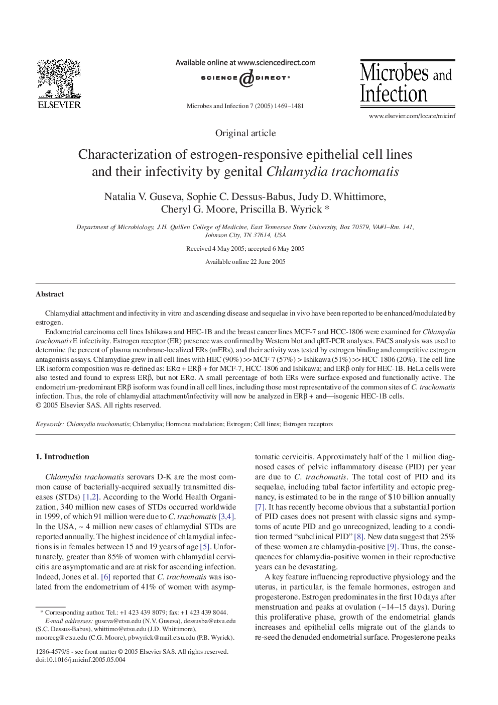
Endometrial carcinoma cell lines Ishikawa and HEC-1B and the breast cancer lines MCF-7 and HCC-1806 were examined for Chlamydia trachomatis E infectivity. Estrogen receptor (ER) presence was confirmed by Western blot and qRT-PCR analyses. FACS analysis was used to determine the percent of plasma membrane-localized ERs (mERs), and their activity was tested by estrogen binding and competitive estrogen antagonists assays. Chlamydiae grew in all cell lines with HEC (90%) >> MCF-7 (57%) > Ishikawa (51%) >> HCC-1806 (20%). The cell line ER isoform composition was re-defined as: ERα + ERβ + for MCF-7, HCC-1806 and Ishikawa; and ERβ only for HEC-1B. HeLa cells were also tested and found to express ERβ, but not ERα. A small percentage of both ERs were surface-exposed and functionally active. The endometrium-predominant ERβ isoform was found in all cell lines, including those most representative of the common sites of C. trachomatis infection. Thus, the role of chlamydial attachment/infectivity will now be analyzed in ERβ + and-isogenic HEC-1B cells.
Related Topics
Life Sciences
Immunology and Microbiology
Immunology
Authors
Natalia V. Guseva, Sophie C. Dessus-Babus, Judy D. Whittimore, Cheryl G. Moore, Priscilla B. Wyrick,
